. 2018 Feb 27;115(9):2174-2179.
doi: 10.1073/pnas.1713301115. Epub 2018 Feb 12.

Intensity and duration of TCR signaling is limited by p38 phosphorylation of ZAP-70T293 and destabilization of the signalosome

Affiliations

Intensity and duration of TCR signaling is limited by p38 phosphorylation of ZAP-70T293 and destabilization of the signalosome

Maria Letizia Giardino Torchia et al. Proc Natl Acad Sci U S A. .

Abstract

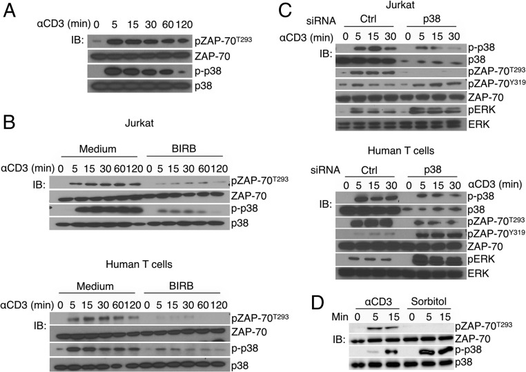
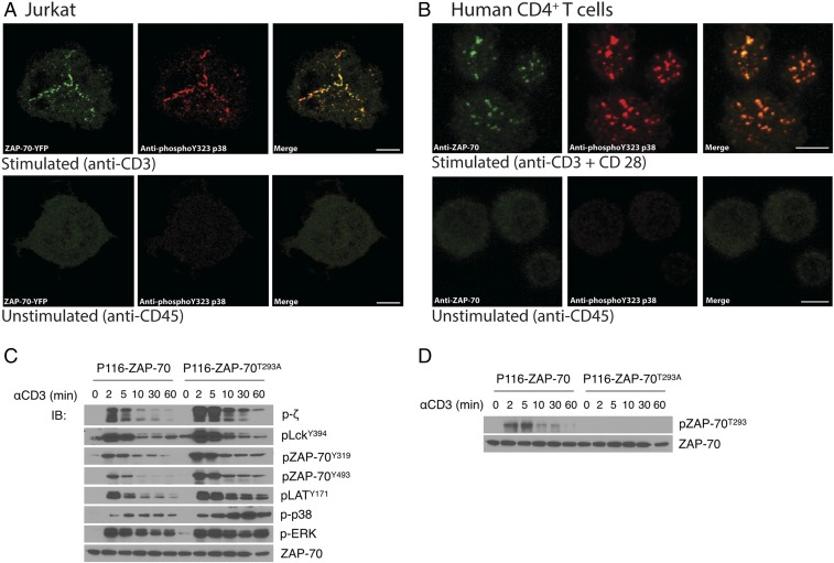
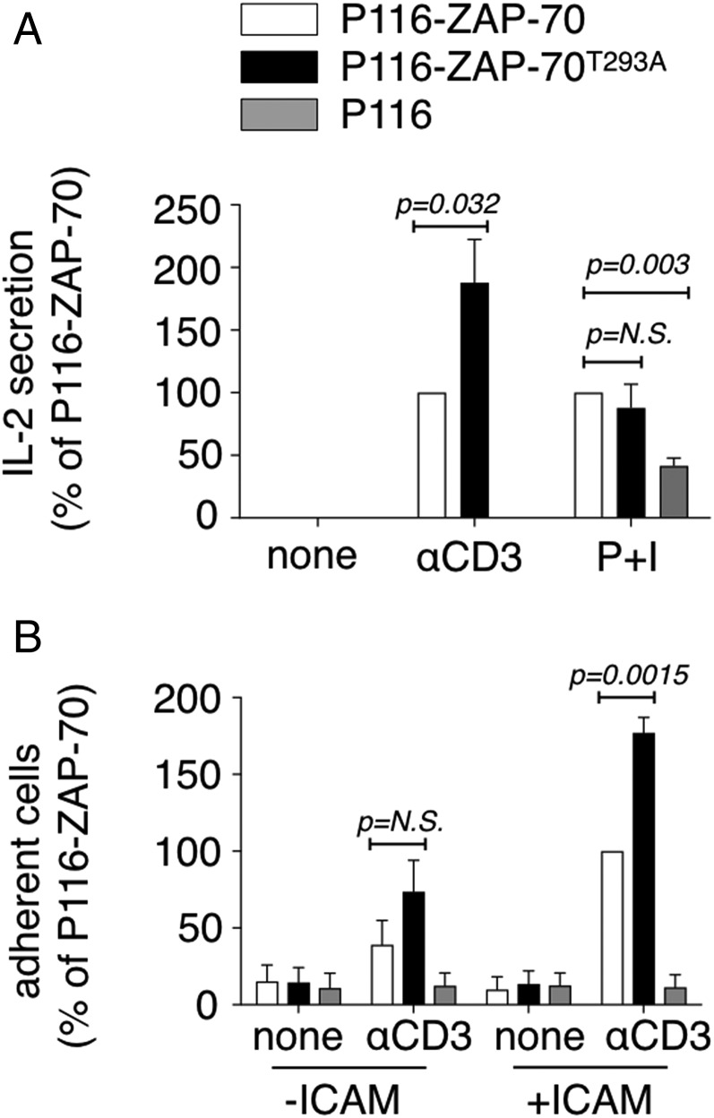
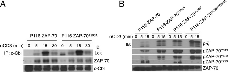
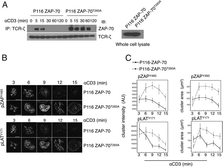
ZAP-70 is a tyrosine kinase that is essential for initiation of T cell antigen receptor (TCR) signaling. We have found that T cell p38 MAP kinase (MAPK), which is directly phosphorylated and activated by ZAP-70 downstream of the TCR, in turn phosphorylates Thr-293 in the interdomain B region of ZAP-70. Mutant T cells expressing ZAP-70 with an alanine substitution at this residue (ZAP-70T293A) had enhanced TCR proximal signaling and increased effector responses. Lack of ZAP-70T293 phosphorylation increased association of ZAP-70 with the TCR and prolonged the existence of TCR signaling microclusters. These results identify a tight negative feedback loop in which ZAP-70-activated p38 reciprocally phosphorylates ZAP-70 and destabilizes the signaling complex.

Keywords: MAP kinase; T cell antigen receptor; immune synapse; signal transduction.


Conflict of interest statement

The authors declare no conflict of interest.

Figures

Fig. 1.
Fig. 1.
p38 phosphorylates ZAP-70T293 in vitro and in T cells activated via the TCR. (A) Jurkat cells were activated with cross-linked anti-CD3 and cell lysates immunoblotted (IB) with antibodies against the indicated proteins. The antibody used to detect p-p38 recognizes both Thr-180/Tyr-182 dual phosphorylation and Thr-180 monophosphorylation (12). (B) Jurkat cells and primary human T cells were activated in the absence or presence of the p38 inhibitor BIRB796 and cell lysates immunoblotted. (C) Validated RNAi to p38α/β were electroporated in Jurkat and primary human T cells, which after 48 were activated with anti-CD3 and analyzed by immunoblotting for the indicated proteins. (D) Jurkat T cells were stimulated with cross-linked anti-CD3 or sorbitol, lysed, and immunoblotted for the indicated proteins. The data are representative of three (A, B, and D) and two (C) independent experiments.
Fig. 2.
Fig. 2.
Phosphorylation of ZAP-70T293 enhances TCR signaling. (A) Jurkat cells stably expressing YFP-tagged ZAP-70 or (B) purified primary human CD4+ T cells were plated on coverslips coated with anti-CD45 (nonactivating) or anti-CD3 (activating) for 3 min, fixed, and immunostained. ZAP-YFP is shown in green and anti-pY323 p38 is shown in red for Jurkat–ZAP-70–YFP cells; ZAP-70 is shown in green and anti-pY323 p38 is shown in red for human T cells. (C and D) ZAP-70–deficient P116 cells expressing WT ZAP-70 or ZAP-70T293A were stimulated with cross-linked anti-CD3 for the indicated times and cell lysates immunoblotted for the indicated proteins. The data are representative of three (A, C, and D) and two (B) independent experiments.
Fig. 3.
Fig. 3.
Phosphorylation of ZAP-70T293 inhibits effector functions. (A) ZAP-70–deficient P116 cells or p116 cells expressing WT ZAP-70 or ZAP-70T293A were stimulated with plate-bound anti-CD3 or PMA and ionomycin (P+I) for 24 h and IL-2 was measured in the supernatant. The data represent the average of three independent experiments and were normalized to the amount of IL-2 produced by P116 cells expressing WT ZAP-70 in each experiment. N.S., not significant. (B) The same cells used in A were activated with cross-linked anti-CD3 and seeded on ICAM1-coated plates for 30 min. Adherent cells were recovered and counted. The data represent the average of five independent experiments and were normalized to binding of P116 cells expressing WT ZAP-70 to iCAM-1 in each experiment.
Fig. 4.
Fig. 4.
Phosphorylation of ZAP-70T293 acts independently of ZAP-70Y292 phosphorylation. (A) ZAP-70–deficient P116 cells expressing WT ZAP-70 or ZAP-70T293A were activated with cross-linked anti-CD3, and cell lysates prepared at the indicated times were immunoprecipitated (IP) with anti–c-Cbl. The immunoprecipitates were resolved by SDS/PAGE and immunoblotted (IB) with antibodies against the indicated proteins. (B) The indicated stable P116 transfectants were stimulated and analyzed as in A. The data are representative of three (A) and two (B) independent experiments.
Fig. 5.
Fig. 5.
Phosphorylation of ZAP-70T293 diminishes recruitment of ZAP-70 to the TCR and destabilizes the signaling complex. (A) ZAP-70–deficient P116 cells expressing WT ZAP-70 or ZAP-70T293A were activated with cross-linked anti-CD3 and cell lysates prepared at the indicated times were immunoprecipitated with anti–TCR-ζ. The immunoprecipitates were resolved by SDS/PAGE and immunoblotted with antibodies against the indicated proteins. The data are representative of three independent experiments. (B, Upper) Representative images illustrating the time course of phosphorylated ZAP-70 clustering in activated Jurkat cells. The images shown here were taken from different staining experiments. (B, Lower) Representative images illustrating the time course of phosphorylated LAT clustering in activated Jurkat cells. The images shown in the Upper and Lower panels were taken from different staining experiments. (C) Quantification of the area of pZAP-70 and pLAT clusters. Error bars show SEM. For WT ZAP-70 cells, 3 min n = 51, 6 min n = 55, 9 min n = 52, 12 min n = 55, and 15 min n = 60. For ZAP-70T293A cells, 3 min n = 55, 6 min n = 50, 9 min n = 52, 12 min n = 53, and 15 min n = 52, taken from three experiments. AU, arbitrary units.

Similar articles

Cited by

References

    1. Smith-Garvin JE, Koretzky GA, Jordan MS. T cell activation. Annu Rev Immunol. 2009;27:591–619. - PMC - PubMed
    1. Acuto O, Di Bartolo V, Michel F. Tailoring T-cell receptor signals by proximal negative feedback mechanisms. Nat Rev Immunol. 2008;8:699–712. - PubMed
    1. Yan Q, et al. Structural basis for activation of ZAP-70 by phosphorylation of the SH2-kinase linker. Mol Cell Biol. 2013;33:2188–2201. - PMC - PubMed
    1. Wang H, et al. ZAP-70: An essential kinase in T-cell signaling. Cold Spring Harb Perspect Biol. 2010;2:a002279. - PMC - PubMed
    1. Hivroz C, Fischer A. Immunodeficiency diseases. Multiple roles for ZAP-70. Curr Biol. 1994;4:731–733. - PubMed

Publication types

MeSH terms

Substances