. 2018 Feb 27;115(9):2192-2197.
doi: 10.1073/pnas.1718144115. Epub 2018 Feb 12.

Epitope-specific monoclonal antibodies to FSHβ increase bone mass

Affiliations

Epitope-specific monoclonal antibodies to FSHβ increase bone mass

Yaoting Ji et al. Proc Natl Acad Sci U S A. .

Abstract

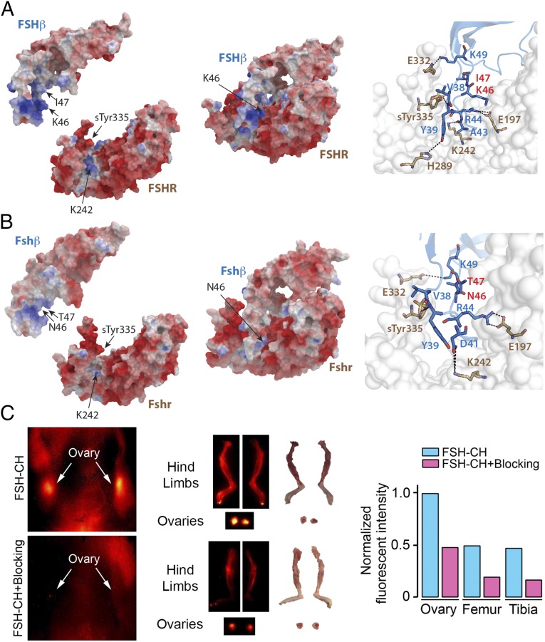
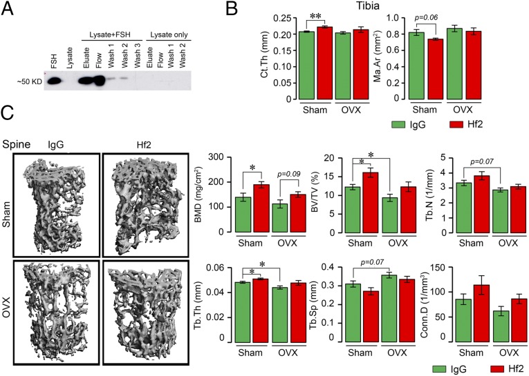
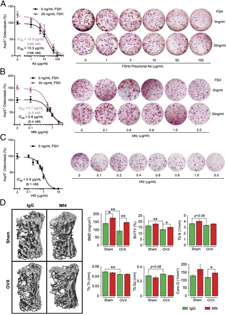
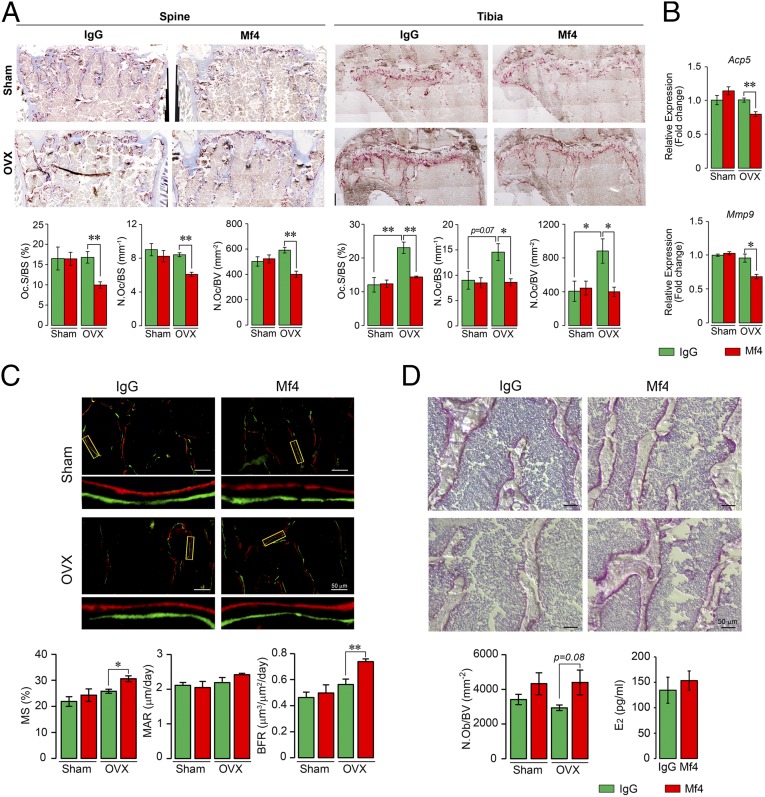
Pituitary hormones have long been thought solely to regulate single targets. Challenging this paradigm, we discovered that both anterior and posterior pituitary hormones, including FSH, had other functions in physiology. We have shown that FSH regulates skeletal integrity, and, more recently, find that FSH inhibition reduces body fat and induces thermogenic adipose tissue. A polyclonal antibody raised against a short, receptor-binding epitope of FSHβ was found not only to rescue bone loss postovariectomy, but also to display marked antiobesity and probeiging actions. Questioning whether a single agent could be used to treat two medical conditions of public health importance--osteoporosis and obesity--we developed two further monoclonal antibodies, Hf2 and Mf4, against computationally defined receptor-binding epitopes of FSHβ. Hf2 has already been shown to reduce body weight and fat mass and cause beiging in mice on a high-fat diet. Here, we show that Hf2, which binds mouse Fsh in immunoprecipitation assays, also increases cortical thickness and trabecular bone volume, and microstructural parameters, in sham-operated and ovariectomized mice, noted on microcomputed tomography. This effect was largely recapitulated with Mf4, which inhibited bone resorption by osteoclasts and stimulated new bone formation by osteoblasts. These effects were exerted in the absence of alterations in serum estrogen in wild-type mice. We also reconfirm the existence of Fshrs in bone by documenting the specific binding of fluorescently labeled FSH, FSH-CH, in vivo. Our study provides the framework for the future development of an FSH-based therapeutic that could potentially target both bone and fat.

Keywords: FSH monoclonal antibody; FSH polyclonal antibody; FSH receptor; antiobesity; osteoporosis treatment.


Conflict of interest statement

The authors declare no conflict of interest.

Figures

Fig. 1.
Fig. 1.
Interaction between the binding domain of human and mouse FSHβ and the FSHR. (A) The crystal structure of FSHβ in complex with the entire ectodomain of FSHR (FSHα omitted for clarity) was used as the template (PDB ID code 4AY9) for comparative modeling. (Right) Dashed lines show interactions between specific amino acids of the human FSH–FSHR complex (discussed in Results). Of note is that K46 and I47 are noninteracting residues. (B) High sequence identity between mouse FSHR (UniProt ID code Q9QWV8) and human FSHR (88%) permitted accurate homology modeling (Modeler, PROCHECK, and PROSA). The FSHβ binding epitope differs by only two amino acids (LVYKDPARPKIQK → LVYKDPARPNTQK). Several models of the mouse Fshβ epitope were constructed (ICM software) (38); restrained minimization was used to remove steric clashes, and the final model was selected on basis of the lowest Cα rmsd after superimposition on template structure (0.2 Å). Fshβ binds in the groove generated between the palm and the thumb of the Fshr. The electrostatic surfaces generate a complementary surface charge (red: acidic residues; blue: basic residues) between the Fshr and Fshβ. Since the crystal structure provides a static snapshot, a short burst of MD simulation was run to allow the side chains of Fshr and Fshβ to dynamically equilibrate and adapt to the contours of the complex (Methods). (Right) Dashed lines show interactions between specific amino acids of the FSH–FSHR complex. N46 and T47 are noninteracting residues. (C) NIR-II imaging of Fshr-expressing ovaries and bones using FSH-CH in adult female mice. FSH-CH (12.5 µg) was injected into the tail vein of mice before in vivo imaging 2 h later. (Left) Supine view showing fluorescent signals in ovaries (Top), which were blocked with 30-fold excess of unconjugated FSH (Bottom); this indicated hormonal specificity of signals. (Middle) NIR-II fluorescence was detected in dissected ovaries and hind limbs (Top); 30-fold excess of unconjugated FSH blocked these signals (Bottom). (Right) Normalized fluorescence intensity for each tissue is shown.
Fig. 2.
Fig. 2.
Monoclonal anti-epitope FSHβ antibody Hf2 binds mouse Fsh to increase bone mass in ovariectomized and sham-operated mice. (A) Recombinant mouse Fsh (Fshα-Fshβ chimera, 2 µg) was added to lysates of HEK293 cells. Spiked and unspiked lysates were passed through resin (Pierce Co-Immunoprecipitation Kit, 26149; Thermo Scientific) with immobilized polyclonal Fsh antibody. Elution (Eluate), flow-through (Flow), and consecutive wash fractions (Wash) were collected and immunoblotted, as shown, with the monoclonal Fsh antibody Hf2. An immunoreactive band at the expected size, ∼50 kDa, in both elution and flow-through fractions is seen in the spiked, but not in unspiked lysate. Lanes 1 and 2 were loaded with mouse FSH and lysate alone, respectively. (B and C) Mice were ovariectomized or sham-operated and injected with Hf2 (raised against YCYTRDLVYKDPARPKIQKTCT) or mouse IgG (200 µg/d) for 4 wk while on normal chow (n = 5–6 mice per group). The vertebral column, and in cases, the long bones were dissected and processed for micro-CT measurements. Cortical bone parameters, namely Ct.Th and marrow area (Ma.Ar), obtained on the middiaphysis of the tibia are shown (B). Representative micro-CT images and structural parameters of vertebral column (spine), namely BMD, BV/TV, Tb.Th, Tb.N, Tb.Sp, and/or Conn.D, are shown (C). Statistics: mean ± SEM; two-tailed Student’s t test, corrected for multiple comparisons; *P ≤ 0.05, **P ≤ 0.01, or as indicated.
Fig. 3.
Fig. 3.
Monoclonal anti-epitope FSHβ antibody increases bone mass in ovariectomized mice. (AC) To confirm FSH specificity of the monoclonal antibodies Mf4 and Hf2, Ficoll-purified hematopoietic stem cells were cultured with RANKL (30 ng/mL) and incubated with varying concentrations of the polyclonal FSH antibody (A), Mf4 (raised against CLVYKDPARPNTQKV) (B), or Hf2 (C) for 5 d in the presence or absence of mouse Fsh (30 ng/mL) (n = 8 wells per group). Cells stained using a kit for tartrate-resistant acid phosphatase (Acp5) were counted. Representative culture wells are shown, together with mean percent osteoclast number (±SEM). Note that the increase in osteoclastogenesis noted with 30 ng/mL FSH (and zero-dose antibody) was abolished progressively with increasing antibody concentrations. IC50s for the polyclonal FSH antibody, Mf4 and Hf2 are shown. (D) Mice were ovariectomized or sham-operated and injected with Mf4 or mouse IgG (100 µg/d) for 4 wk while on normal chow (n = 5 mice per group). The vertebral column was dissected and processed for micro-CT measurements. Representative micro-CT images and structural parameters, namely BMD, BV/TV, Tb.Th, Tb.N, Tb.Sp, and/or Conn.D, are shown. Statistics: mean ± SEM; two-tailed Student’s t test, corrected for multiple comparisons; *P ≤ 0.05, **P ≤ 0.01, or as shown.
Fig. 4.
Fig. 4.
Monoclonal anti-epitope FSHβ antibody Mf4 reduces bone resorption and stimulates osteoblastic bone formation. The vertebral column (spine) and tibia from the mice above (Fig. 3C) were dissected and processed for Acp5 staining. Shown are representative images of Acp5-stained sections from vertebral column (spine) and tibial head (tibia), with parameters of resorption, namely Oc.S/BS, N.Oc/BS, and N.Oc/BV (A). mRNA expression (qPCR) for Acp5 and Mmp9 in marrow-derived osteoclasts obtained from these mice showed reduced osteoclastogenesis in the Mf4-treated group (B). These mice were also injected with calcein and xylelol orange 5 d apart before sacrifice, and bones were processed for dynamic histomorphometry and alkaline phosphatase staining of osteoblasts. Shown are fluorescent micrographs of vertebral column displaying xylelol orange (red) and calcein (green) labels, and estimates of MS, MAR, or interlabel distance, and BFR (C). Alkaline phosphatase-labeled sections and calculated N.Ob/BV are shown (D). Statistics: n = 5 mice per group; for B, n = 3, technical replicates; mean ± SEM; two-tailed Student’s t test, corrected for multiple comparisons; *P ≤ 0.05, **P ≤ 0.01, or as shown.

References

    1. Greendale GA, Sowers M. The menopause transition. Endocrinol Metab Clin North Am. 1997;26:261–277. - PubMed
    1. Sowers MR, et al. Endogenous hormones and bone turnover markers in pre- and perimenopausal women: SWAN. Osteoporos Int. 2003;14:191–197. - PubMed
    1. Sowers MR, et al. Changes in bone resorption across the menopause transition: Effects of reproductive hormones, body size, and ethnicity. J Clin Endocrinol Metab. 2013;98:2854–2863. - PMC - PubMed
    1. Randolph JF, Jr, et al. The value of follicle-stimulating hormone concentration and clinical findings as markers of the late menopausal transition. J Clin Endocrinol Metab. 2006;91:3034–3040. - PubMed
    1. Sowers M, Pope S, Welch G, Sternfeld B, Albrecht G. The association of menopause and physical functioning in women at midlife. J Am Geriatr Soc. 2001;49:1485–1492. - PubMed

Publication types

Substances