. 2018 Feb 13;9(1):e02381-17.
doi: 10.1128/mBio.02381-17.

The Birth and Demise of the IS Apl1- mcr-1-IS Apl1 Composite Transposon: the Vehicle for Transferable Colistin Resistance

Affiliations

The Birth and Demise of the IS Apl1- mcr-1-IS Apl1 Composite Transposon: the Vehicle for Transferable Colistin Resistance

Erik Snesrud et al. mBio. .

Abstract

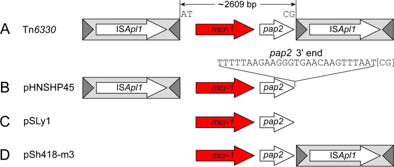
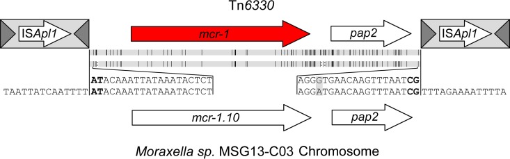
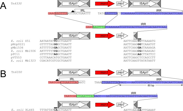
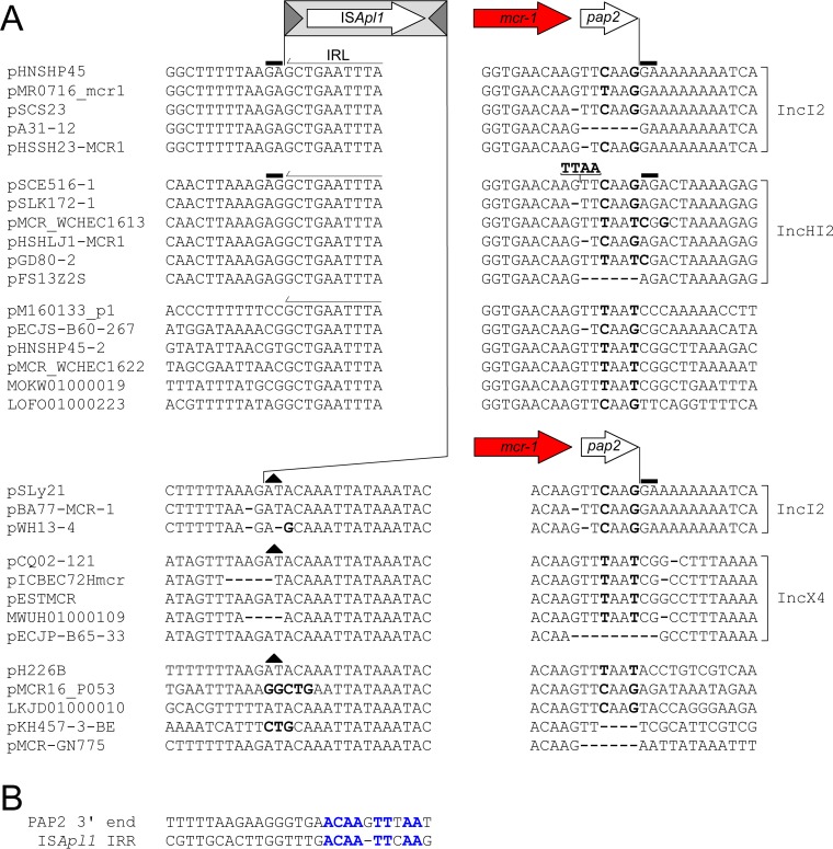
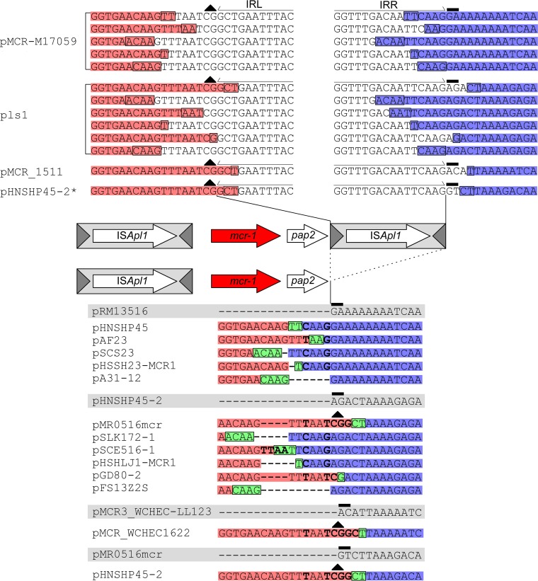
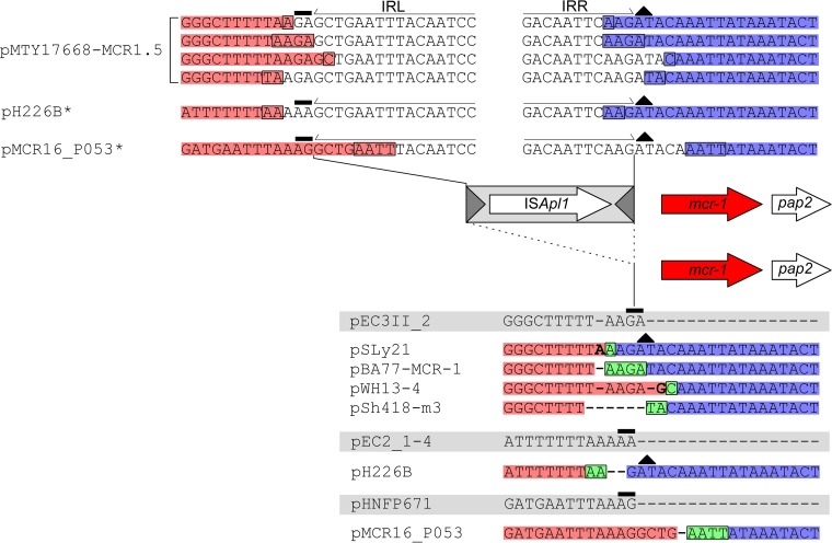
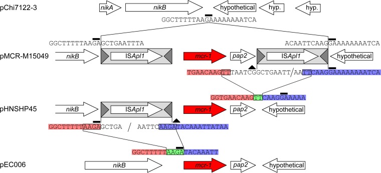
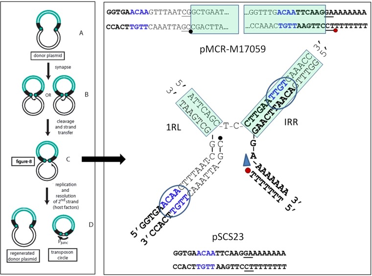
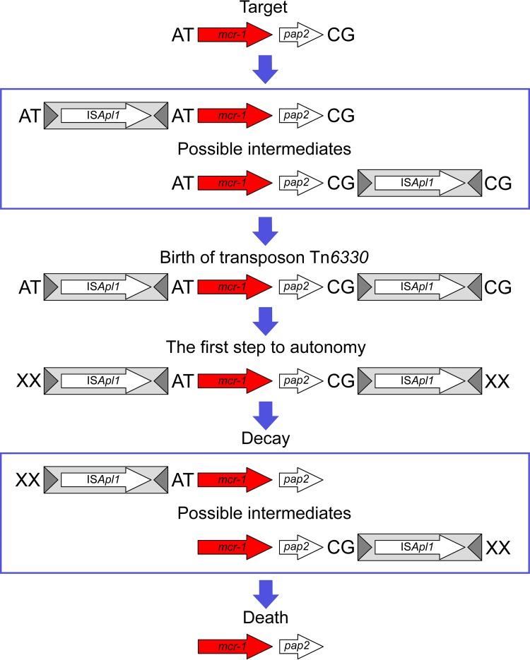
The origin and mobilization of the ~2,609-bp DNA segment containing the mobile colistin resistance gene mcr-1 continue to be sources of uncertainty, but recent evidence suggests that the gene originated in Moraxella species. Moreover mcr-1 can be mobilized as an ISApl1-flanked composite transposon (Tn6330), but many sequences have been identified without ISApl1 or with just a single copy (single ended). To further clarify the origins and mobilization of mcr-1, we employed the Geneious R8 software suite to comprehensively analyze the genetic environment of every complete mcr-1 structure deposited in GenBank as of this writing (September 2017) both with and without associated ISApl1 (n = 273). This revealed that the 2,609-bp mcr-1 structure was likely mobilized from a close relative of a novel species of Moraxella containing a chromosomal region sharing >96% nucleotide identity with the canonical sequence. This chromosomal region is bounded by AT and CG dinucleotides, which have been described on the inside ends (IE) of all intact Tn6330 described to date and represent the ancestral 2-bp target site duplications (TSDs) generated by ISApl1 transposition. We further demonstrate that all mcr-1 structures with just one ISApl1 copy or with no ISApl1 copies were formed by deletion of ISApl1 from the ancestral Tn6330, likely by a process related to the "copy-out-paste-in" transposition mechanism. Finally, we show that only the rare examples of single-ended structures that have retained a portion of the excised downstream ISApl1 including the entire inverted right repeat might be capable of mobilization.IMPORTANCE A comprehensive analysis of all intact mcr-1 sequences in GenBank was used to identify a region on the chromosome of a novel Moraxella species with remarkable homology to the canonical mcr-1 structure and that likely represents the origin of this important gene. These data also demonstrate that all mcr-1 structures lacking one or both flanking ISApl1 were formed from ancestral composite transposons that subsequently lost the insertion sequences by a process of abortive transposition. This observation conclusively shows that mobilization of mcr-1 occurs as part of a composite transposon and that structures lacking the downstream ISApl1 are not capable of mobilization.

Keywords: colistin resistance; composite transposon formation; drug resistance evolution; insertion sequence; transposon decay.


Figures

FIG 1
FIG 1
Four representative sequences showing the four general mcr-1 structures identified to date. (A) The composite transposon Tn6330 (13). (B) pHNSHP45, a single-ended structure with an upstream copy of ISApl1 only. (C) pSLy1, a structure lacking both copies of ISApl1. (D) pSh418-m3, a single-ended structure with a downstream copy of ISApl1 only. The ~2,609-bp region comprising the mcr-1 region is shown flanked by the conserved, ancestral TSD dinucleotides AT and CG on the IE. The last 27 bp of the mcr-1 region that comprise the 3′ end of the putative pap2 gene are indicated for clarity. ISApl1, light gray box; transposase, long white arrow; terminal inverted repeats, dark gray triangle; mcr-1, red arrow; pap2, short white arrow. The same scheme is used for all of the figures.
FIG 2
FIG 2
Alignment of Tn6330 and the homologous region on the Moraxella sp. MSG13-CO3 chromosome. The consensus sequence at the end of both regions is provided, with the conserved AT and CG dinucleotides that are found on the IE of all Tn6330 highlighted in bold. Mutations and deletions are indicated by vertical black lines and colons, respectively, between the two images.
FIG 3
FIG 3
Schematic representation of single-ended mcr-1 cassettes that have retained the last 42 bp (A) or 90 bp (B) of an ancestral ISApl1 that includes the entire IRR. The conserved, ancestral CG dinucleotide on the IE of the downstream ISApl1 is indicated with a black triangle. The bases upstream and downstream of the deletion that are retained after ISApl1 loss are highlighted in red and blue, respectively. The deletion joints upstream and downstream of the ISApl1 are encased in a black rectangle. The bases upstream and downstream of the deletion that are retained after ISApl1 loss are highlighted in red and blue, respectively, while the remaining copy of the deletion joint that is retained after the two ends are joined following ISApl1 excision is highlighted in green and encased in a black rectangle. The same labeling scheme is used throughout the figures. Putative 2-bp TSDs in 5 of the 7 sequences are highlighted in bold.
FIG 4
FIG 4
(A) Consensus alignment of multiple mcr-1 regions from a variety of plasmid backbones with one (single-ended) flanking copy or no flanking copy of ISApl1. The minor sequence variation at the 3′ end of the pap2 gene in all structures and at the 5′ end of mcr-1 in structures lacking the upstream ISApl1 is highlighted in bold text, with absent bases indicated by bold dashes. To preserve the alignment, the additional TTAA tetranucleotide in pSCE516-1 has been placed above the sequence, with a line indicating the correct position. (B) Alignment of the last 27 bp of the pap2 gene and the 26 bp that constitute the IRR of ISApl1. The nucleotides that form the basis for the sequence variability noted in structures that have lost the downstream ISApl1 are highlighted in blue.
FIG 5
FIG 5
Alignment showing the decay of multiple different instances of Tn6330 (top) into the corresponding single-ended structures formed (bottom of figure), aligned with their respective empty sites (bottom of figure, highlighted in gray; see the text for more details). “Hypothetical” Tn6330 insertions constructed from known empty site plasmids, the corresponding single-ended structures, and the sequence of Tn6330 are indicated with an asterisk. The labeling scheme is identical to that described for Fig. 3.
FIG 6
FIG 6
Alignment showing the decay of multiple different instances of Tn6330 (top) into the corresponding double-deletion structures formed (bottom of figure), aligned with their respective empty sites (below, highlighted in gray; see the text for details). “Hypothetical” Tn6330 insertions constructed from known empty site plasmids, the corresponding double-deletion structures, and the sequence of Tn6330 are indicated with an asterisk. Dashes were inserted to maintain sequence alignment.
FIG 7
FIG 7
Alignment showing the formation and successive decay of Tn6330 in a set of four highly related IncI2 plasmids. The labeling scheme is identical to that described for Fig. 3.
FIG 8
FIG 8
A mechanism for ISApl1 deletion. The left hand panel shows part of the ISApl1 transposition cycle. The IS is shown in green and the donor plasmid backbone in black. The terminal IRs are indicated by black boxes and the tips of the IS as green circles. (A to C) The IS ends in the donor plasmid (A) undergo synapsis (B), and one or other end undergoes cleavage to generate a 3′OH which then attacks the opposite end at a position several nucleotides into the donor backbone to generate a molecule in a figure eight formation in which the two IRs are joined by a single-strand bridge (C). (D) A 3′OH generated on the donor plasmid is then used to replicate the IS, generating a double-strand circular transposition intermediate. A strong promoter is generated by the juxtaposition of the two IS ends which drives high levels of transposase expression, facilitating insertion into a suitable target DNA (not shown). The right panel (top) shows the double-strand sequence of the IS ends in IncI2 plasmid pMCR-M17059 as an example. The middle panel presents the structure of the single-strand bridged molecule (as described for panel C) that is shown in the left panel. IS ends are boxed in green. The 3′OH generated in the donor plasmid DNA is indicated by a red dot and the corresponding 5′ phosphate at the other IS end by a black dot. The blue arrow indicates the direction of transposition-associated replication. The deletion joint is shown in blue. The sequence remaining after deletion (bottom) representing plasmid pSCS23 is composed of the bold black characters together with one of the blue tetranucleotide sequences.
FIG 9
FIG 9
The birth and demise of Tn6330. A schematic representation of the birth of Tn6330 following insertion of two copies of ISApl1 into the chromosome of a species closely resembling Moraxella sp. MSG13-C03 followed by successive decay of the transposon due to the loss of one ISApl1 copy and then both ISApl1 copies is shown. The conserved AT and CG dinucleotides that were formed during the first sequestration of the mcr-1 region, and that are now found on the IE of all instances of Tn6330, are highlighted in bold.

Similar articles

Cited by

References

    1. Watkins RR, Bonomo RA. 2016. Overview: global and local impact of antibiotic resistance. Infect Dis Clin North Am 30:313–322. doi:10.1016/j.idc.2016.02.001. - DOI - PubMed
    1. Siguier P, Gourbeyre E, Varani A, Ton-Hoang B, Chandler M. 2014. Everyman’s guide to bacterial insertion sequences, p 555–590. In Craig NL, Chandler M, Gellert M, Lambowitz AM, Rice PA, Sandmeyer SB (ed), Mobile DNA III, 3rd ed. ASM Press, Washington DC.
    1. Nagy Z, Chandler M. 2004. Regulation of transposition in bacteria. Res Microbiol 155:387–398. doi:10.1016/j.resmic.2004.01.008. - DOI - PubMed
    1. Machida Y, Machida C, Ohtsubo E. 1982. A novel type of transposon generated by insertion element IS102 present in a pSC101 derivative. Cell 30:29–36. doi:10.1016/0092-8674(82)90008-3. - DOI - PubMed
    1. Poirel L, Decousser JW, Nordmann P. 2003. Insertion sequence ISEcp1B is involved in expression and mobilization of a bla(CTX-M) beta-lactamase gene. Antimicrob Agents Chemother 47:2938–2945. doi:10.1128/AAC.47.9.2938-2945.2003. - DOI - PMC - PubMed

Publication types

MeSH terms

LinkOut - more resources