. 2018 Feb 13;8(1):2924.
doi: 10.1038/s41598-018-20608-6.

Type 2 Innate Lymphocytes Actuate Immunity Against Tumours and Limit Cancer Metastasis

Affiliations

Type 2 Innate Lymphocytes Actuate Immunity Against Tumours and Limit Cancer Metastasis

Iryna Saranchova et al. Sci Rep. .

Abstract

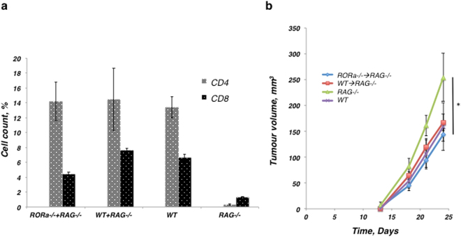
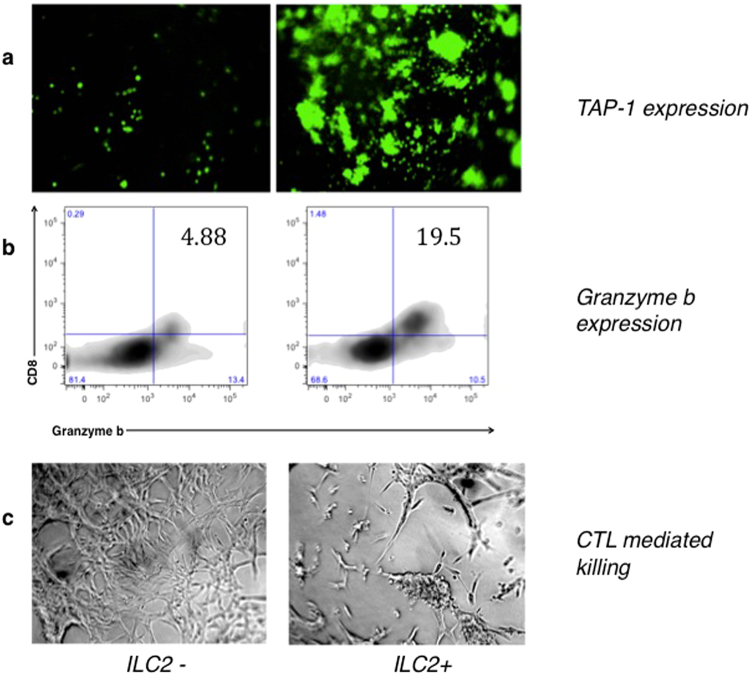
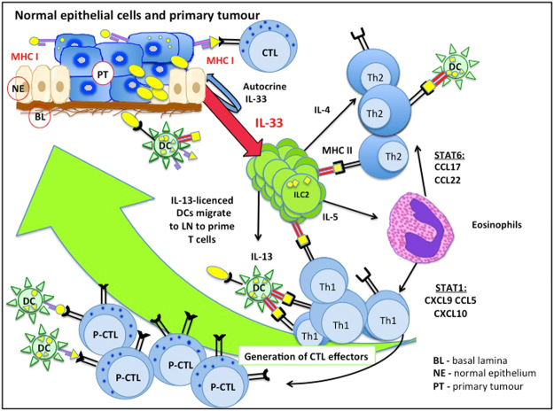
Type 2 innate lymphoid cells (ILC2) potentiate immune responses, however, their role in mediating adaptive immunity in cancer has not been assessed. Here, we report that mice genetically lacking ILC2s have significantly increased tumour growth rates and conspicuously higher frequency of circulating tumour cells (CTCs) and resulting metastasis to distal organs. Our data support the model that IL-33 dependent tumour-infiltrating ILC2s are mobilized from the lungs and other tissues through chemoattraction to enter tumours, and subsequently mediate tumour immune-surveillance by cooperating with dendritic cells to promote adaptive cytolytic T cell responses. We conclude that ILC2s play a fundamental, yet hitherto undescribed role in enhancing anti-cancer immunity and controlling tumour metastasis.


Conflict of interest statement

The authors declare no competing interests.

Figures

Figure 1
Figure 1
The study models of antecedent tumour cell lines, TC1/A9, retain their immunological characteristics both in vitro and in vivo. (a) Flow cytometry shows the MHC-I expression levels of tumour cells grown in vitro, where the primary cells (TC1) express higher levels of MHC-I than their metastatic derivatives (A9). (b,c) Resected tumour sections were stained with antibodies against MHC-I. (b) Primary TC1 tumours retain MHC-I expression in vivo. (c) The corresponding metastatic A9 tumour has greatly reduced MHC-I expression. (d,e) Morphology of tumours is shown with H&E staining. (d) Primary TC1 tumours resemble a sponge structure that clearly distinguishes them from (e) metastatic A9 tumours, which appear uniformly distributed, with a solid architecture. 10 µm thick sections were imaged at 20x (b,c) or 4x (d,e) magnification. Size bar = 100 µm in (b,c); Size bar = 500 µm in (d,e).
Figure 2
Figure 2
Genetic complementation of immune evasive tumours shows a clear phenotypic shift towards immune recognition. Immunohistochemical staining was used to visualise tumour infiltrating immune cells. CD4 is a glycoprotein found on the surface of T helper cells, macrophages, dendritic cells. CD8 is primarily a marker for cytotoxic T cells, but also found on natural killer cells and DCs; CD68 is a marker of monocytes and macrophages; Ly6G is a marker for neutrophils; FoxP3 is a marker of regulatory T cells. Greater numbers of CD4+ and CD8+ cells can be seen within (c,g) the genetically modified (metastatic A9+IL-33) tumours versus unmodified metastatic (A9) tumours (a,e). Fewer regulatory T cells are present in IL-33 expressing tumours, as indicated by lower FoxP3 staining; metastatic A9 (q) versus metastatic A9+IL-33 (s) or primary TC1 (r) tumours. Increased macrophage and neutrophil responses are seen in IL-33 expressing tumours: unmodified metastatic A9 (i,m) versus metastatic A9+IL-33 (k,o) or primary TC1 (j,n) tumours respectively. (d,h,l,p,t) Negative controls (rat IgG targeting Keyhole Limpet Hemocyanin (KLH)) were included to show that non-specific staining was minimized. The percentage of positively stained cells was calculated from the total number of cells on each slide. 10 µm thick sections were stained with appropriate antibodies and imaged at 20X magnification (at); size bar = 100 µm (at).
Figure 3
Figure 3
The frequency of ILC2s is elevated in primary tumours and metastatic tumours expressing IL-33, when compared to metastatic tumours not expressing IL-33. (a) Gating strategy: ILC2s from tumours were sorted by FACS as LinST2+CD127+CD90.2+ cells. A total of 2 × 105 cellular events from 1 g of disaggregated tumours were used to create a profile for each tumour. The ILC2 detection strategy included: gating on lineage-negative (Lin) and Thy1.2-positive lymphocytes, with further analysis for ST2+ and CD127+ expression. The final gate (double positive for ST2 and CD127) indicates ILC2s isolated and sorted from tumour tissue, as a percentage of the LinThy1.2+ cells isolated. Note, EGFP-positive tumour cells corresponded to the FITC-labeled-Lin+ gate and thus also appear in the “Lin+” gate, however these cells are not positive for Thy1.2, ST2 or CD127 and are therefore removed by subsequent gates. (b) Isolated ILC2s appeared to be fully functional and retained the ability to secrete IL-13 and IL-5. This graph shows ILC2s, which were isolated from either disaggregated draining lymph nodes (LN), including mesenteric, inguinal and lumbar LNs, or from TC1 tumours. Tumour cells (TC1) do not secret IL-5 and IL-13 and were used as negative controls for interleukin secretion levels. The ranges represent the data from animals within each tumour group, where lymph nodes (n = 8 animals) and tumours (n = 4 animals). (c) The percentage of ILC2 cells isolated from total cells in the tumour is represented in the bar graph. The number of ILC2s that could be isolated from the tumour went up in direct relation to the ability of the tumour cells to secrete IL-33. This difference was statistically significant between the number of ILC2 cells isolated from the primary TC1 and metastatic A9 tumours (*P < 0.05; Student’s t-test). Error bars represent standard error of the mean.
Figure 4
Figure 4
The mice lacking ILC2s (RORα−/−) were less able to limit the growth of tumours. Tumours with and without IL-33-expression were established in the WT and RORα−/− chimeric mice. (a) The presence of ILC2s significantly inhibited tumour formation in WT chimeric mice bearing tumours expressing IL-33, when compared to RORα−/− chimeras, which lacked ILC2s. Metastatic A9 tumours demonstrated rapid progression and severe disease symptoms reaching the humane end-point that resulted in early termination of animals from both A9 chimeric groups, indicating that the ILC2s were not significantly able to control the growth of fast growing tumours which lack IL-33 expression. Metastatic A9+IL-33 tumour growth was more aggressive in RORα−/− chimeric mice lacking ILC2s, to the point where they resembled the growth of A9 cells alone. The difference in growth results between metastatic A9+IL-33 tumours grown in WT versus RORα−/− chimeric mice was significant; P < 0.002 (Student t-test). Primary TC1 tumours were able to grow faster in mice lacking ILC2s (RORα−/− chimeras) than in WT chimeras P < 0.05 (Student t-test). (b) The numbers of ILC2 cells found in neighbouring lymph nodes were significantly lower in RORα−/− mice compared to WT chimeras bearing IL-33 expressing tumours. This served as a quality control for bone marrow transplantation; *P < 0.05 (Student t-test). (c) The numbers of ILC2 cells found in primary TC1 tumours were significantly higher than in metastatic A9 or A9+IL-33 tumours; *P < 0.05 (Student t-test). (d,e) RORα deficiency had no effect on the percentage of CD4+ or CD8+ lymphocytes found in either lymph nodes or tumours in response to (d) primary TC1 or (e) metastatic A9+IL-33 tumours. The percentage of all cells was calculated as a fraction of 2 × 105 cellular events used to create a profile for each organ or tissue. The error bars represent standard error of the mean; n = 8 mice per group.
Figure 5
Figure 5
Functional ILC2 cells are important in controlling the metastatic spread of the tumours. (a) Spread of EGFP-positive circulating tumour cells (CTC) to distal organs was higher in chimeric mice lacking ILC2s (RORα−/− mice); even primary TC1 cells that are not metastatic in the WT mice could be found beyond the initial site of transplantation in mice lacking ILC2s. (b) In lungs, CTC counts were higher in animals with reduced number of ILC2s (RORα−/−). (c) The number of ILC2s present in the lungs appeared to go up with the presence of CTCs in the lungs. The percentage of all cells was calculated from the fraction of live cells in 2 × 105 events used to create a profile for each organ; n = 8 mice per group; *P < 0.05; **P < 0.01.
Figure 6
Figure 6
RORα−/− mice produce functional lymphocytes. (a) Immune cell count in the peripheral blood of (WT → RAG−/−) and (RORα−/− → RAG−/−) animals, as well as in the WT and RAG−/− control animals. (b) The T and B cells isolated from the RORα−/− and WT mice were equivalent in their functional ability to suppress the tumour growth to the level of the WT control group, however, the tumour growth was significantly higher in RAG−/− control group when compared to the (RORα−/− → RAG−/−) group of mice (*P = 0.29, Student t-test). The percentage of all cells was calculated as a fraction of 2 × 105 cellular events used to create a profile for each organ or tissue. The error bars represent standard error of the mean; n = 8 mice per group.
Figure 7
Figure 7
Impact of ILC2s on specific CTL effector mechanisms. Co-culture of metastatic murine prostate TAP-1-low carcinoma cells (LMD) and CD8 T cells with ILC2s (right panel) or without ILC2 cells (left panel): (a) TAP-1 expression level in LMD cells after activation with ILC2 cells (right) and without ILC2 cells (left); (b) Granzyme b expression by CTL cells in the absence of ILC2s (left) or in the presence of ILC2s (right); (c) Light microscope images of cell co-cultures after the incubation with CTLs show the extent of CTL-mediated killing, either in the presence or absence of ILC2s.
Figure 8
Figure 8
Heat map illustrates the relative expression of genes in metastatic A9+vector cells, metastatic A9+IL-33 cells and primary TC1+vector cells. The Mouse Cancer Inflammation & Immunity Crosstalk RT2 Profiler PCR Array was used to analyze cDNA from the tumour cell lines grown in vitro. Relative changes in gene expression were calculated using the ΔΔCt (threshold cycle) method. Changes in cycle threshold (ΔCt) values for each tested gene were obtained by subtracting the average of the threshold cycle (Ct) values for the housekeeping genes (Gusb, Hsp90ab1, Gapdh, and Actβ) from the threshold cycle value of the gene. The expression fold change was calculated as (2(−ΔΔCt)). Expression of the housekeeping genes between the tumour cell samples (metastatic A9+vector; metastatic A9+IL-33; primary TC1+vector) were normalized to the transcription levels seen in the metastatic A9+vector samples. The ratio up- or down-regulation was then calculated relative to the normalized transcription of each gene in each of the tumour cell lines. (a) Immunostimulatory factors are down-regulated in metastatic cells (A9), whereas factors involved into immunosuppressive pathways are reduced in primary tumours (TC1). Antigen presenting components are over expressed in primary tumours that is well in line with up-regulation of chemo-attractants and with a better anti-tumour immune response. (b) The level of CXCL12 expression is almost 1000-fold higher in IL-33 expressing primary tumour cells (TC1) and metastatic cells (A9+IL-33) when compared to metastatic tumour cells (A9 alone), P < 0.01 (Student t-test).
Figure 9
Figure 9
Model linking the adaptive and innate immune responses during tumour development via IL-33-ILC2 axis. IL-33-expressing tumour environment stimulates the development of ILC2 cells and functionally activates them through the ST2 receptor pathway. Functionally active ILC2s alter the tumour microenvironment triggering both innate and adaptive immune responses. ILC2s recruit DCs through IL-13 production, stimulate Th2 cells and possibly Th1 cells through direct ILC2 antigen presentation via MHC-II molecules, and indirectly stimulate CTL precursor cells through DC endogenous antigen presentation or cross-presentation via MHC-I molecules. Th1 and Th2 cells may also be activated by DCs through exogenous antigen presentation via MHC-II molecules. Through the release of IL-5 by ILC2s and subsequent recruitment of eosinophils, the chemokine profiles of tumour microenvironment is changed to attract Th1 and CD8+ T cells and to direct the activation of CTL-mediated killing and cancer rejection. This mechanism acts to suppress the frequency of circulating tumours cell and subsequent metastasis. In metastatic tumours with low IL-33 content, the IL-33/ILC2 pathway is not initiated and immune escape is facilitated.

References

    1. Lanier LL. Shades of grey–the blurring view of innate and adaptive immunity. Nat Rev Immunol. 2013;13:73–74. doi: 10.1038/nri3389. - DOI - PubMed
    1. Vivier E, et al. Innate or adaptive immunity? The example of natural killer cells. Science. 2011;331:44–49. doi: 10.1126/science.1198687. - DOI - PMC - PubMed
    1. Chan IH, et al. Interleukin-23 is sufficient to induce rapid de novo gut tumorigenesis, independent of carcinogens, through activation of innate lymphoid cells. Mucosal Immunol. 2014;7:842–856. doi: 10.1038/mi.2013.101. - DOI - PubMed
    1. Kirchberger S, et al. Innate lymphoid cells sustain colon cancer through production of interleukin-22 in a mouse model. J Exp Med. 2013;210:917–931. doi: 10.1084/jem.20122308. - DOI - PMC - PubMed
    1. Shields JD, Kourtis IC, Tomei AA, Roberts JM, Swartz MA. Induction of lymphoidlike stroma and immune escape by tumors that express the chemokine CCL21. Science. 2010;328:749–752. doi: 10.1126/science.1185837. - DOI - PubMed

Publication types