. 2018 Feb 13;8(1):2952.
doi: 10.1038/s41598-018-21087-5.

Vascular dysfunction in obese diabetic db/db mice involves the interplay between aldosterone/mineralocorticoid receptor and Rho kinase signaling

Affiliations

Vascular dysfunction in obese diabetic db/db mice involves the interplay between aldosterone/mineralocorticoid receptor and Rho kinase signaling

Aurelie Nguyen Dinh Cat et al. Sci Rep. .

Abstract

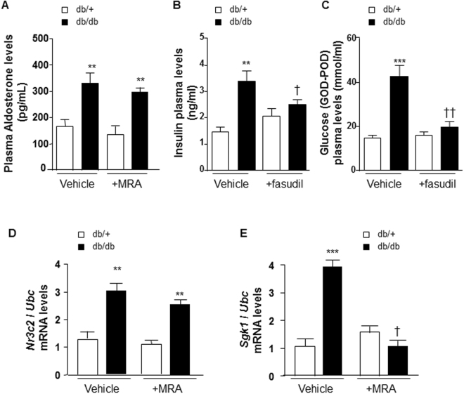
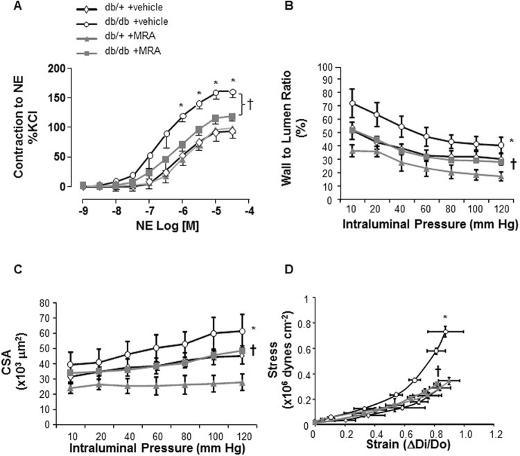
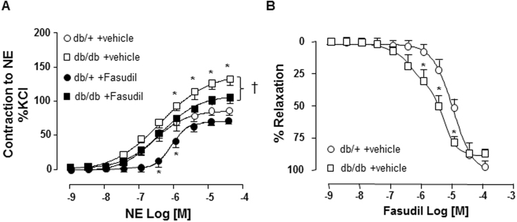
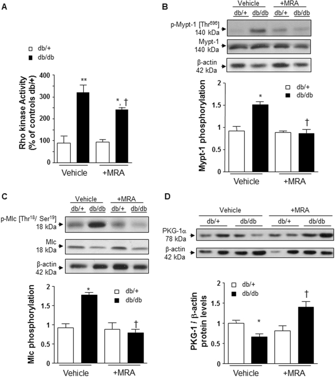
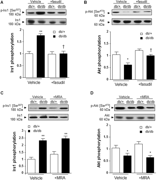
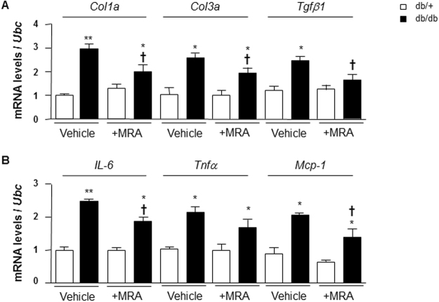
Activation of aldosterone/mineralocorticoid receptors (MR) has been implicated in vascular dysfunction of diabetes. Underlying mechanisms are elusive. Therefore, we investigated the role of Rho kinase (ROCK) in aldosterone/MR signaling and vascular dysfunction in a model of diabetes. Diabetic obese mice (db/db) and control counterparts (db/+) were treated with MR antagonist (MRA, potassium canrenoate, 30 mg/kg/day, 4 weeks) or ROCK inhibitor, fasudil (30 mg/kg/day, 3 weeks). Plasma aldosterone was increased in db/db versus db/+. This was associated with enhanced vascular MR signaling. Norepinephrine (NE)-induced contraction was increased in arteries from db/db mice. These responses were attenuated in mice treated with canrenoate or fasudil. Db/db mice displayed hypertrophic remodeling and increased arterial stiffness, improved by MR blockade. Vascular calcium sensitivity was similar between depolarized arteries from db/+ and db/db. Vascular hypercontractility in db/db mice was associated with increased myosin light chain phosphorylation and reduced expression of PKG-1α. Vascular RhoA/ROCK signaling and expression of pro-inflammatory and pro-fibrotic markers were exaggerated in db/db mice, effects that were attenuated by MRA. Fasudil, but not MRA, improved vascular insulin sensitivity in db/db mice, evidenced by normalization of Irs1 phosphorylation. Our data identify novel pathways involving MR-RhoA/ROCK-PKG-1 that underlie vascular dysfunction and injury in diabetic mice.


Conflict of interest statement

The authors declare no competing interests.

Figures

Figure 1
Figure 1
Plasma aldosterone, insulin and glucose concentrations and Nr3c2 and Sgk1 levels in arteries from db/db versus db/+ mice. (A) Plasma aldosterone levels are increased in db/db. MR antagonist treatment has no effect. Non fasting plasma levels of insulin (B) and glucose (C) are increased in db/db vs db/+ mice, and fasudil reduces both in db/db, but not in db/+. Nr3c2 (D) and Sgk1 (E) mRNA levels are increased in mesenteric arteries from db/db vs db/+ mice. MR antagonist treatment abolished increased Sgk1, but not Nr3c2 mRNA levels in mesenteric arteries from db/db versus db/+ mice. Ubc was used as housekeeping gene for normalization. Results are expressed mean ± SEM, n = 6–8 mice per group, **p < 0.01, ***p < 0.001 db/db vs db/+, p < 0.05, ††p < 0.01 vehicle vs +MRA. MRA: MR antagonist (canrenoate); Nr3c2: nuclear receptor subfamily 3 group C member 2 (mineralocorticoid receptor); vs: versus. Vehicle: saline NaCl 0.9%; Ubc: ubiquitin C.
Figure 2
Figure 2
Vascular remodeling and function in arteries from db/db versus db/+ mice. (A) NE-induced contraction of mesenteric resistance arteries from control db/+ and db/db mice, treated by an MR antagonist (30 mg/kg/day, 4 weeks) or vehicle (saline), was performed using a wire myograph. Increased NE-induced contractility in db/db mice was prevented by canrenoate treatment. Structural and mechanical parameters were assessed by pressurized myography in mesenteric arteries from db/db vs db/+ mice. Vascular wall to lumen ratio (B) and CSA (C) were increased in db/db mice. (D) The stress-strain relationship was shifted to the left indicating that vascular stiffness is increased in db/db vs db/+ mice. All of these vascular changes were prevented by canrenoate treatment. Results are expressed mean ± SEM, n = 6–8 mice/group, *p < 0.05 db/db vs db/+, p < 0.05 vehicle vs +MRA. CSA: cross sectional area; MRA: MR antagonist (canrenoate). Vehicle: saline NaCl 0.9%.
Figure 3
Figure 3
Rho kinase inhibitor fasudil improved contractility of arteries from db/db mice. (A) NE-induced contraction of mesenteric resistance arteries from control db/+ and db/db mice, treated by a Rho kinase inhibitor Fasudil (30 mg/kg/day, 3 weeks) or vehicle (saline), was performed using a wire myograph. (B) Dose-dependent fasudil-induced relaxation curve (10−9 to 10−4 mol/l) was shifted to the left for db/db vs db/+ mice. Data are presented as mean ± SEM; n = 6–8 mice/group, *p < 0.05 db/db vs db/+, p < 0.05 vehicle vs +fasudil. NE: norepinephrine; Vehicle: saline NaCl 0.9%.
Figure 4
Figure 4
Rho kinase activity is increased in mesenteric arteries from db/db versus db/+ mice. (A) Rho kinase activity was assessed by enzymatic immunoassay. Upregulation of downstream targets of Rho kinase activation with Mypt-1 (B) and Mlc (C) phosphorylation in db/db vs db/+ mice. (d) PKG-1α protein levels are decreased in mesenteric arteries from db/db vs db/+ mice. These changes were prevented by canrenoate treatment. Results are expressed as mean ± SEM, n = 6–8 mice per group, *p < 0.05, **p < 0.01 db/db vs db/+, p < 0.05 vehicle vs +MRA. Vehicle: saline NaCl 0.9%.
Figure 5
Figure 5
Fasudil improved vascular insulin signaling in db/db versus db/+ mice, and this is MR-independent pathway. Fasudil treatment prevents increased Ser-307-phosphorylation of Irs1 (A) and decreased Ser-473 phosphorylation of Akt (B) in mesenteric arteries from db/db vs db/+ mice. However, canrenoate treatment has no effect on the increase in Irs1 phosphorylation (C) neither on the decrease in Akt phosphorylation (D) in db/db mice. Data are presented as mean ± SEM; n = 6 to 8 mice/group. *p < 0.05, **p < 0.01 db/db vs db/+. Irs1: Insulin receptor substrate 1; MRA: MR antagonist canrenoate. Vehicle: saline NaCl 0.9%. Results are expressed mean ± SEM, n = 6–8 mice per group, *p < 0.05, **p < 0.01 db/db vs db/+, p < 0.05, ††p < 0.01 vehicle vs +fasudil. Irs1: insulin receptor substrate 1; Vehicle: saline NaCl 0.9%.
Figure 6
Figure 6
Pro-fibrotic and pro-inflammatory markers mRNA levels in arteries from db/db and db/+ mice. (A) Pro-fibrotic markers such as Col1a, Col3a and Tgfb1 mRNA levels were increased in mesenteric arteries from db/db compared to db/+ mice. (B) Pro-inflammatory markers such as IL-6, Tnfa and Mcp-1 mRNA levels were increased in mesenteric arteries from db/db compared to db/+ mice. Ubc was used as housekeeping gene for normalization. Results are expressed mean ± SEM, n = 6–8 mice per group, *p < 0.05, **p < 0.01 db/db vs db/+, p < 0.05 vehicle vs +MRA. Vehicle: saline NaCl 0.9%. IL-6: interleukine-6; Mcp-1: monocyte chemoattractant protein 1; Tnfα: tumor necrosis factor alpha; Tgfβ1: transforming growth factor beta 1; Ubc: ubiquitin C.
Figure 7
Figure 7
Hypothetical scheme of putative signaling mechanisms induced by aldosterone/MR over-activation in obesity. On one hand, aldosterone/MR induces ROCK activity through decreased PKG-1a, resulting in vascular hypercontractility of arteries from obese db/db mice. In obesity-related diabetes, ROCK may also contribute to vascular insulin resistance independently of MR. In addition, vascular remodeling, due to increased pro-fibrotic and pro-inflammatory signaling, is also regulated, in part, by MR. MLC: myosin light chain; Mypt1: myosin light chain phosphatase subunit 1; MR: mineralocorticoid receptor; PKG-1α: cGMP-dependent protein kinase G type 1 isoform alpha; ROCK: Rho kinase; p: phosphorylated.

References

    1. Goodfriend TL, Kelley DE, Goodpaster BH, Winters SJ. Visceral obesity and insulin resistance are associated with plasma aldosterone levels in women. Obes Res. 1999;7:355–362. doi: 10.1002/j.1550-8528.1999.tb00418.x. - DOI - PubMed
    1. Rossi GP, et al. Primary Aldosteronism Prevalence in hYpertension Study Investigators. Body mass index predicts plasma aldosterone concentrations in overweight-obese primary hypertensive patients. J Clin Endocrinol Metab. 2008;93:2566–2571. doi: 10.1210/jc.2008-0251. - DOI - PubMed
    1. Guo C, et al. Mineralocorticoid receptor blockade reverses obesity-related changes in expression of adiponectin, peroxisome proliferator-activated receptor-gamma, and proinflammatory adipokines. Circulation. 2008;117:2253–2261. doi: 10.1161/CIRCULATIONAHA.107.748640. - DOI - PMC - PubMed
    1. Nguyen Dinh Cat A, et al. Adipocyte-derived factors regulate vascular smooth muscle cells through mineralocorticoid and glucocorticoid receptors. Hypertension. 2011;58:479–488. doi: 10.1161/HYPERTENSIONAHA.110.168872. - DOI - PubMed
    1. Briones AM, et al. Adipocytes produce aldosterone through calcineurin-dependent signalling pathways: implications in diabetes mellitus-associated obesity and vascular dysfunction. Hypertension. 2012;59:1069–1078. doi: 10.1161/HYPERTENSIONAHA.111.190223. - DOI - PubMed

Publication types

MeSH terms

Substances