Acquired ventricular septal defect due to infective endocarditis
- PMID: 29440841
- PMCID: PMC5803962
- DOI: 10.4103/apc.APC_130_17
Acquired ventricular septal defect due to infective endocarditis
Abstract
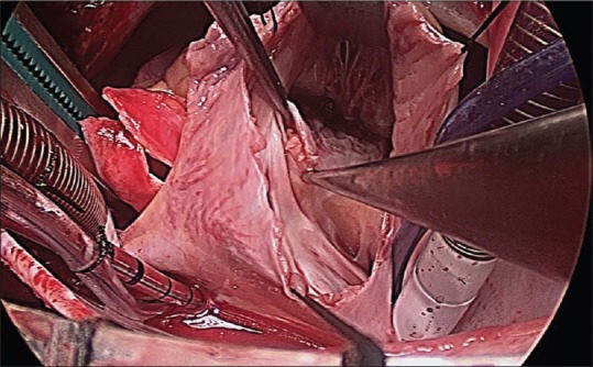
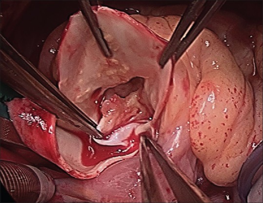
Acquired intracardiac left-to-right shunts are rare occurrences. Chest trauma and myocardial infection are well-known causes of acquired ventricular septal defect (VSD). There have been several case reports describing left ventricle to right atrium shunt after infective endocarditis (IE). We present here a patient found to have an acquired VSD secondary to IE of the aortic and tricuspid valves in the setting of a known bicuspid aortic valve. This is the first case reported of acquired VSD in a pediatric patient in the setting of IE along with literature review of acquired left-to-right shunts.
Keywords: Endocarditis; infective endocarditis; ventricular septal defect.
Conflict of interest statement
There are no conflicts of interest.
Figures


References
-
- Ota T, Yamaguchi R, Tanigawa T, Otuka K, Hayashi Y, Nishiyama H, et al. Left ventricular-right atrial communication by perforation of the atrioventricular portion of the membranous septum and severe aortic valve regurgitation caused by infective endocarditis. J Echocardiogr. 2011;9:30–2. - PubMed
-
- Sinisalo JP, Sreeram N, Jokinen E, Qureshi SA. Acquired left ventricular-right atrium shunts. Eur J Cardiothorac Surg. 2011;39:500–6. - PubMed
-
- Sun LC, Lai CC, Wang CY, Wang YH, Wang JY, Hsu YL, et al. Risk factors for infective endocarditis in children with congenital heart diseases – A nationwide population-based case control study. Int J Cardiol. 2017;248:126–30. - PubMed
-
- Baltimore RS, Gewitz M, Baddour LM, Beerman LB, Jackson MA, Lockhart PB, et al. Infective endocarditis in childhood: 2015 update: A Scientific statement from the american heart association. Circulation. 2015;132:1487–515. - PubMed
-
- Tribak M, Konaté M, Elhassani A, Mahfoudi L, Jaabari I, Elkenassi F, et al. Aortic infective endocarditis: Value of surgery. About 48 cases. Ann Cardiol Angeiol (Paris) 2016;65:15–20. - PubMed
Publication types
Grants and funding
LinkOut - more resources
Full Text Sources
Other Literature Sources