Review
. 2018 Jan 30:12:1.
doi: 10.3389/fnana.2018.00001. eCollection 2018.

Layer- and Cell Type-Specific Modulation of Excitatory Neuronal Activity in the Neocortex

Affiliations
Review

Layer- and Cell Type-Specific Modulation of Excitatory Neuronal Activity in the Neocortex

Gabriele Radnikow et al. Front Neuroanat. .

Abstract

From an anatomical point of view the neocortex is subdivided into up to six layers depending on the cortical area. This subdivision has been described already by Meynert and Brodmann in the late 19/early 20. century and is mainly based on cytoarchitectonic features such as the size and location of the pyramidal cell bodies. Hence, cortical lamination is originally an anatomical concept based on the distribution of excitatory neuron. However, it has become apparent in recent years that apart from the layer-specific differences in morphological features, many functional properties of neurons are also dependent on cortical layer or cell type. Such functional differences include changes in neuronal excitability and synaptic activity by neuromodulatory transmitters. Many of these neuromodulators are released from axonal afferents from subcortical brain regions while others are released intrinsically. In this review we aim to describe layer- and cell-type specific differences in the effects of neuromodulator receptors in excitatory neurons in layers 2-6 of different cortical areas. We will focus on the neuromodulator systems using adenosine, acetylcholine, dopamine, and orexin/hypocretin as examples because these neuromodulator systems show important differences in receptor type and distribution, mode of release and functional mechanisms and effects. We try to summarize how layer- and cell type-specific neuromodulation may affect synaptic signaling in cortical microcircuits.

Keywords: acetylcholine; adenosine; barrel cortex; cortical layers; dopamine; neuromodulation; orexin.


Figures

FIGURE 1
FIGURE 1
Excitatory neuron types in layers 2–6 of the (A) medial prefrontal and (B) primary somatosensory (barrel) cortex. Different excitatory neuron types in cortical layers 2–6 (L2–L6) of rat mPFC and S1 barrel cortex are shown. Most neuron types are pyramidal cells with apical dendrites of different shape and length with the exception of spiny stellate cells in layer 4 and multipolar neurons in layer 6B. Somatodendritic domains are shown in different shades of red, with bright red indicating superficial and dark red deep layers. Note that the diversity of excitatory neurons is much higher than that shown here and that even between e.g., sensory cortices different pyramidal cell types can be found. L2P: L2 pyramidal cell; L3stP: L3 slender-tufted pyramidal cell, L3btP: L3 broad-tufted pyramidal cell; L3P: L3 pyramidal cell; L4SSC: L4 spiny stellate cell; L4SP: L4 star pyramidal cell; L5stP: L5 slender-tufted pyramidal cell (with strong axonal projections to layer 2 and 3); L5utP: L5 untufted pyramidal cell; L5btP: L5 broad-tufted pyramidal cell and L5ttP: L5 thick-tufted pyramidal cell (both of which project mainly to subcortical targets); L6A tall P: L6A tall pyramidal cell; L6A wide P: L6A wide pyramidal cell; L6A invP: L6A inverted pyramidal cell; L6AccP: L6A corticocortical pyramidal cell; L6ActP: L6A corticothalamic pyramidal cellL6AP: L6BP L6B pyramidal cell; L6BMC: L6B multipolar cell. This terminology will be used throughout the remainder of the text.
FIGURE 2
FIGURE 2
Signaling cascades of neuromodulator-coupled G-proteins. Signaling pathways of the G-protein coupled receptors (GPCRs) discussed in this review (top row). (A) Gi/o signaling, (B) Gs signaling and (C) Gq/11 signaling pathways. Signaling occurs via the dissociated and phosphorylated Gα subunit or via direct interaction between the βγ subunit complex and the effector (K+- and Ca2+ channels). See text for details. It should be noted that the downstream signaling pathways of PKA, PKC and PLC are significantly more diverse than shown here. (AC, adenylate cyclase; ADP, adenosine diphosphate; ATP, adenosine trisphosphate; cAMP, cyclic adenosine monophosphate; DAG, diacylglycerol; GIRK, G-protein coupled, inwardly rectifying K+ channel; IP3, inositol trisphosphate; PKA, phosphokinase A; PKC, phosphokinase C; PLC; phospholipase C). For the abbreviation of receptor subtypes see text.
FIGURE 3
FIGURE 3
Layer- and cell type-specific difference in the adenosine response in the somatosensory barrel cortex. Significant layer- and cell type-specific differences in the adenosine response in cortical layers 2, 5A and 5B (A) Reconstructions of L2, slender-tufted L5A and thick-tufted L5B pyramidal cells in the in the somatosensory barrel cortex. (B) Voltage response to adenosine application in L2 and L5A pyramidal cells; L2 pyramidal cells are almost unresponsive to adenosine. (C) Comparison of the adenosine response in layers 2, 5A and 5B showing layer-specific differences in the amplitude of the hyperpolarization. This may indicate cell type-specific differences in the density of adenosine A1ARs. L3, L4, and L6 pyramidal cells show also a hyperpolarizing response to adenosine (not shown). Modified from van Aerde et al. (2015).
FIGURE 4
FIGURE 4
Layer- and cell type-specific A1 adenosine receptor distribution in the prefrontal and primary somatosensory barrel cortex. Adenosine receptors on excitatory neocortical neurons can be found in cortical layers 3–6. Note that in both prefrontal and somatosensory cortex L2 (upper L2/3) pyramidal cells with broad apical tufts were unresponsive to adenosine suggesting no or a very low expression of A1 adenosine receptors. In layer 5 of somatosensory cortex two pyramidal cell types showed marked differences in their adenosine response that was correlated with their morphology and laminar location; such a clear difference was not found for the prefrontal cortex. All data are from rat.
FIGURE 5
FIGURE 5
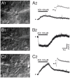
Layer- and cell type-specific muscarinic effects of acetylcholine in the somatosensory barrel cortex. Layer–specific response of excitatory neurons in S1 barrel cortex to rapid application of ACh. (A1–C1) Differential interference contrast images of the recorded neurons in layers 2/3, 4 and 5; the profile of the solution ejected by the puff pipette is outlined in white dotted lines. (A2–C2) Example responses of L2/3, L4 and L5 excitatory neurons to puff application of ACh. Pyramidal cells in layer 2/3 (A) and 5 (C) show a depolarization in response to ACh (duration indicated by bar) that is sometimes preceded by a transient hyperpolarization (gray trace in inset). In contrast, all L4 excitatory neurons show a persistent and monophasic hyperpolarizing ACh response.
FIGURE 6
FIGURE 6
Layer-specific nAChR responses in pyramidal cells of the PFC. Current responses of L2/3, L5, and L6 PFC pyramidal cells to rapid ACh application. (A) AP firing pattern elicited by 300 ms current steps in PFC pyramidal cells of cortical layers 2/3, 5, and 6. (B) Morphological reconstructions of example L2/3, L5, and L6 pyramidal cells, in green, black and red, respectively. The recording (left) and puff pipette for rapid application) are shown at the soma of the L2/3 pyramidal cell (C) Response of pyramidal cells to brief applications of ACh. About 90% of L2/3 pyramidal cells did not display a nicotinergic ACh response (top trace). A small fraction (∼10%) of L2/3 and all L5 pyramidal cells showed rapid inward currents following ACh application, a hallmark of α7 nAChR-mediated currents. L6 pyramidal cells showed very slowly desensitizing ACh-induced currents that are mediated by α4β2α5 nAChRs (see text for details). After Poorthuis et al. (2013a) with permission from Oxford University Press.
FIGURE 7
FIGURE 7
Expression of mAChRs and nAChRs in the neocortex. Schematic diagram of the layer- and cell-type specific distribution of nAChRs and mAChRs in the neocortex. Cortical layering is indicated on the left. Pyramidal cells (PC) in layers 2/3, 5A, 5B and 6 are shown; L5A are generally slender-tufted and L5B thick-tufted pyramidal cells. L4 excitatory neurons (L4 ExcN) include L4 spiny stellate, star pyramids and pyramidal cells. The different brain regions from which the mAChR and nAChR distribution were obtained are given in brackets.
FIGURE 8
FIGURE 8
Expression of dopamine receptors in the neocortex. Schematic diagram of the layer- and cell-type specific distribution of dopamine receptor types in different pyramidal cell types in the neocortex. Data were obtained for pyramidal cells (PC) in layers 2/3, 5, and 6; CC and CT denote L5 PC with corticocortical and corticothalamic projection targets. L4 excitatory neurons (L4 ExcN) include L4 spiny stellate, star pyramids and pyramidal cells. Brain regions for which the receptor distribution were obtained are given in brackets. Apart from L4 ExcN all data are from functional, mainly electrophysiological studies (see text for details).

References

    1. Alexandre C., Andermann M. L., Scammell T. E. (2013). Control of arousal by the orexin neurons. Curr. Opin. Neurobiol. 23 752–759. 10.1016/j.conb.2013.04.008 - DOI - PMC - PubMed
    1. Alves N. C., Bailey C. D., Nashmi R., Lambe E. K. (2010). Developmental sex differences in nicotinic currents of prefrontal layer VI neurons in mice and rats. PLOS ONE 5:e9261. 10.1371/journal.pone.0009261 - DOI - PMC - PubMed
    1. Ariano M. A., Sibley D. R. (1994). Dopamine receptor distribution in the rat CNS: elucidation using anti-peptide antisera directed against D1A and D3 subtypes. Brain Res. 649 95–110. 10.1016/0006-8993(94)91052-9 - DOI - PubMed
    1. Arroyo S., Bennett C., Aziz D., Brown S. P., Hestrin S. (2012). Prolonged disynaptic inhibition in the cortex mediated by slow, non-alpha7 nicotinic excitation of a specific subset of cortical interneurons. J. Neurosci. 32 3859–3864. 10.1523/JNEUROSCI.0115-12.2012 - DOI - PMC - PubMed
    1. Astman N., Gutnick M. J., Fleidervish I. A. (1998). Activation of protein kinase C increases neuronal excitability by regulating persistent Na+ current in mouse neocortical slices. J. Neurophysiol. 80 1547–1551. 10.1152/jn.1998.80.3.1547 - DOI - PubMed

LinkOut - more resources