. 2018 Jan 30:9:25.
doi: 10.3389/fphar.2018.00025. eCollection 2018.

15, 16-Dihydrotanshinone I Inhibits Hemangiomas through Inducing Pro-apoptotic and Anti-angiogenic Mechanisms in Vitro and in Vivo

Affiliations

15, 16-Dihydrotanshinone I Inhibits Hemangiomas through Inducing Pro-apoptotic and Anti-angiogenic Mechanisms in Vitro and in Vivo

Yihong Cai et al. Front Pharmacol. .

Abstract

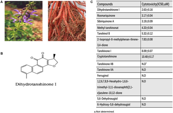
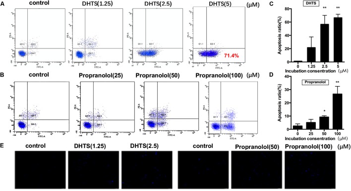
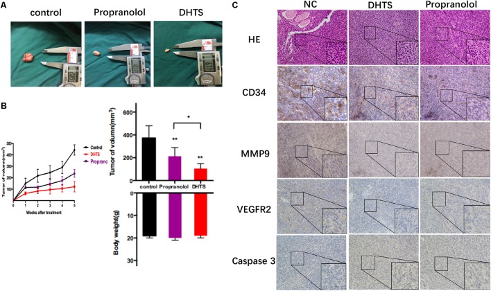
Infantile hemangioma (IH) is a common and benign vascular neoplasms, which has a high incidence in children. Although IH is benign, some patients experience complications such as pain, functional impairment, and permanent disfigurement. Treatment options for IH include corticosteroids, surgery, vincristine, interferon or cyclophosphamide. However, none of these modalities are ideal due to restrictions or potential serious side effects. There is thus a great need to explore novel treatments for IH with less side effects. Angiogenesis, vasculogenesis and tumorigenesis are the main features of IH. Tanshen is mostly used in Chinese traditional medicine to treat hematological abnormalities. Therefore, the aim of our study was to evaluate anti-proliferation and anti-angiogenesis effects on hemangiomas cells by extracted Tanshen compounds compared with propranolol, the first-line treatment for IH currently, both in vitro and in vivo. Cell viability, apoptosis, protein expression and anti-angiogenesis were analyzed by CCK8, Annexin V staining, Western blot and tube formation, respectively. The anti-tumor activity in vivo was evaluated using a mouse xenograft model. Fourteen major compounds extracting from Tanshen were screened for their ability to inhibit hemangiomas cells. Of the 14 compounds investigated, 15,16-Dihydrotanshinone I (DHTS) was the most potent modulator of EOMA cell biology. DHTS could significantly decrease EOMA cells proliferation by inducing cell apoptosis, which is much more efficient than propranolol in vitro. DHTS increased the expression of several apoptosis-related proteins, including caspase9, caspase3, PARP, AIF, BAX, cytochrome c, caspase8 and FADD and significantly inhibited angiogenesis, as indicated by reduced tube formation and diminished expression of vascular endothelial cell growth factor receptor 2 and matrix metalloproteinase 9. In nude mice xenograft experiment, DHTS (10 mg/kg) could significantly inhibit the tumor growth of EOMA cells as well as propranolol (40 mg/kg). Our study showed that DHTS was much more effective than propranolol in inhibiting hemangiomas proliferation and angiogenesis in vitro and in vivo, which could have potential therapeutic applications for treatment of IH.

Keywords: 15,16-dihydrotanshinone I; anti-angiogenesis; apoptosis; infantile hemangiomas; propranolol.


Figures

FIGURE 1
FIGURE 1
The cytotoxity of extracted fourteen compounds of Tanshen on EOMA. (A) The pictures of Tanshen. (B) Chemical structures of Dihydrotanshinone I. (C) Half inhibition concentration (IC50) of extracted Tanshen on EOMA cells.
FIGURE 2
FIGURE 2
15,16-Dihydrotanshinone I (DHTS) and propranolol inhibited cell proliferation. (A) DHTS inhibited the proliferation of EOMA cell lines dose-dependently as well as propranolol. Cell viability determined by CCK8 assay. IC50 value treated by DHTS was 2.63 ± 0.16 μM, while 53.6 ± 1.73 μM by propranolol. (B) Images of cell colonies after treatment with different concentrations of DHTS or propranolol for 24 h. (C) Morphologic characteristics of EOMA cells which treated by DHTS, propranolol or DMSO (control) were observed by microscopy.
FIGURE 3
FIGURE 3
15,16-Dihydrotanshinone I and propranolol induced time-dependent apoptosis in EOMA cells. (A,B) EOMA cells were treated with 2.5 μM DHTS and 50 μM propranolol for 0, 24, or 48 h. Cells were collected and were detected with flow cytometry analysis. (C,D) The quantitative data showed the percentage of apoptotic cells in (A,B). P < 0.05, ∗∗P < 0.01 compared with drug-untreated group.
FIGURE 4
FIGURE 4
15,16-Dihydrotanshinone I and propranolol induced dose-dependent apoptosis in EOMA cells. (A,B) EOMA cells were treated with different concentrations of propranolol (0, 25, 50, and 100 μM) and DHTS (0, 1.25, 2.5, and 5 μM). Cells were collected and were detected with flow cytometry analysis. (C,D) The quantitative data showed the percentage of apoptotic cells in A and B. P < 0.05, ∗∗P < 0.01 compared with drug-untreated group; (E) The Hochest33342 staining assay revealed that DHTS and propranolol facilitated cell apoptosis in EOMA cells.
FIGURE 5
FIGURE 5
Apoptosis related proteins were detected in EOMA cells after treated with DHTS and propranolol. (A,B) DHTS induced PARP, Aif, Caspase9, Caspase3, Bax and Cyts3 in low concentration more than in high concentration, while induced FADD and Caspase 8 more in relatively high concentration. (C,D) Propranolol induced PARP, Aif, Caspase9, Caspase3, Bax and Cyts3 in high concentration more than in low concentration, while induced FADD and Caspase 8 more in relatively low concentration.
FIGURE 6
FIGURE 6
Schematic diagram of DHTS-induced mitochondria- and Fas-mediated apoptosis.
FIGURE 7
FIGURE 7
Effects of DHTS and propranolol on tube formation of EOMA cells. (A,B) Cell cultured on Matrigel, treated by DHTS with different concentrations (0, 1.25, and 2.5 μM) after 4 h and then photographed under a microscope. P < 0.05, ∗∗P < 0.01 compared with normal group. (C,D) Cell cultured on Matrigel and treated by propranolol (0, 25, and 50 μM) with different concentrations after 4h. P < 0.05, ∗∗ P < 0.01 compared with normal group. (E,F) VEGFR2 and MMP9 were both down-regulated by DHTS and propranolol treatment.
FIGURE 8
FIGURE 8
15,16-Dihydrotanshinone I and propranolol inhibits tumor growth in vivo. (A) The tumors were excised from mice incubated EOMA cells after treatment and measured by digital caliper. Propranolol and DHTS inhibited tumor growth after treatment. (B) Tumor volumes were measured by digital caliper three times a week. Figures showed tumor and body weights of mice after final treatment. ∗∗P < 0.01 (One-Way ANOVA was used for the data analysis) compared with control group, P>0.05, The plotted error bars represent mean ± SEM. (C) CD34, MMP9, VEGFR2 and Caspase3 were detected by Immunohistochemistry after xenograft tumor tissues were excised.

References

    1. Albiñana V., Poveda L., Zarrabeitia R., Bernabéu C., Botella L. M. (2012). Propranolol as antiangiogenic candidate for the therapy of hereditary haemorrhagic telangiectasia. Thromb. Haemost. 108 41–53. 10.1160/TH11-11-0809 - DOI - PubMed
    1. Albiñana V., Villar Gómez de Las Heras K., Serrano-Heras G., Segura T., Perona-Moratalla A. B., Mota-Pérez M. (2015). Propranolol reduces viability and induces apoptosis in hemangioblastoma cells from von Hippel-Lindau patients. Orphanet J. Rare Dis. 10:118. 10.1186/s13023-015-0343-5 - DOI - PMC - PubMed
    1. Atalay M., Gordillo G., Roy S., Rovin B., Bagchi D., Bagchi M., et al. (2003). Anti-angiogenic property of edible berry in a model of hemangioma. FEBS Lett. 544 252–257. 10.1016/S0014-5793(03)00509-X - DOI - PubMed
    1. Chang J., Most D., Bresnick S., Mehrara B., Steinbrech D. S., Reinisch J., et al. (1999). Proliferative hemangiomas: analysis of cytokine gene expression and angiogenesis. Plast. Reconstr. Surg. 103 1–9. 10.1097/00006534-199901000-00001 - DOI - PubMed
    1. Chang L. C., Haggstrom A. N., Drolet B. A., Baselga E., Chamlin S. L., Garzon M. C., et al. (2008). Growth characteristics of infantile hemangiomas: implications for management. Pediatrics 122 360–367. 10.1542/peds.2007-2767 - DOI - PubMed