. 2018 Feb 14;14(2):e1007223.
doi: 10.1371/journal.pgen.1007223. eCollection 2018 Feb.

The PHD finger protein Spp1 has distinct functions in the Set1 and the meiotic DSB formation complexes

Affiliations

The PHD finger protein Spp1 has distinct functions in the Set1 and the meiotic DSB formation complexes

Céline Adam et al. PLoS Genet. .

Abstract

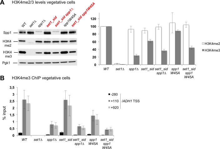
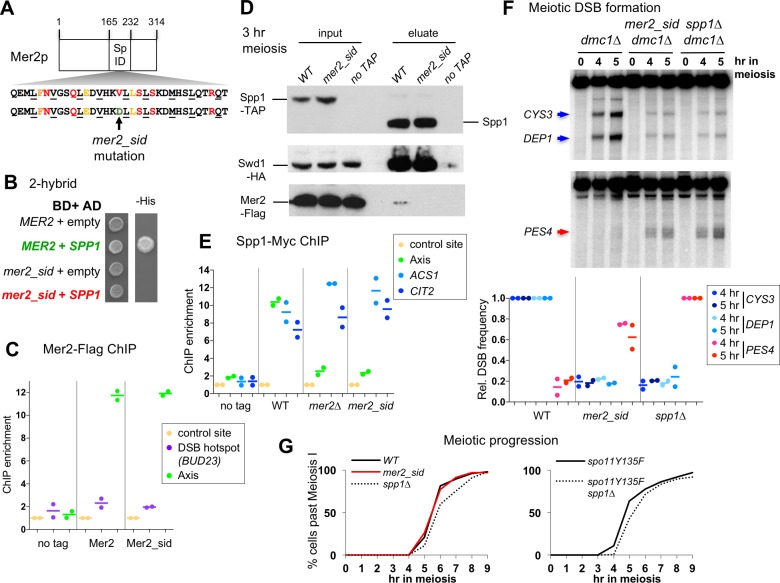
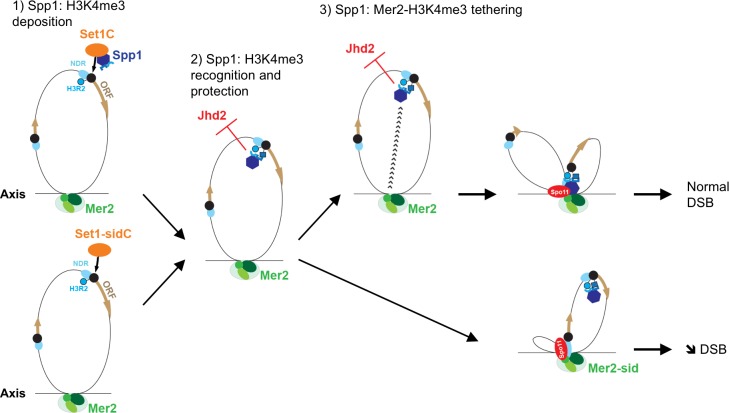
Histone H3K4 methylation is a feature of meiotic recombination hotspots shared by many organisms including plants and mammals. Meiotic recombination is initiated by programmed double-strand break (DSB) formation that in budding yeast takes place in gene promoters and is promoted by histone H3K4 di/trimethylation. This histone modification is recognized by Spp1, a PHD finger containing protein that belongs to the conserved histone H3K4 methyltransferase Set1 complex. During meiosis, Spp1 binds H3K4me3 and interacts with a DSB protein, Mer2, to promote DSB formation close to gene promoters. How Set1 complex- and Mer2- related functions of Spp1 are connected is not clear. Here, combining genome-wide localization analyses, biochemical approaches and the use of separation of function mutants, we show that Spp1 is present within two distinct complexes in meiotic cells, the Set1 and the Mer2 complexes. Disrupting the Spp1-Set1 interaction mildly decreases H3K4me3 levels and does not affect meiotic recombination initiation. Conversely, the Spp1-Mer2 interaction is required for normal meiotic recombination initiation, but dispensable for Set1 complex-mediated histone H3K4 methylation. Finally, we provide evidence that Spp1 preserves normal H3K4me3 levels independently of the Set1 complex. We propose a model where Spp1 works in three ways to promote recombination initiation: first by depositing histone H3K4 methylation (Set1 complex), next by "reading" and protecting histone H3K4 methylation, and finally by making the link with the chromosome axis (Mer2-Spp1 complex). This work deciphers the precise roles of Spp1 in meiotic recombination and opens perspectives to study its functions in other organisms where H3K4me3 is also present at recombination hotspots.


Conflict of interest statement

The authors have declared that no competing interests exist.

Figures

Fig 1
Fig 1. Spp1 interacts with the Set1 complex and the Mer2 DSB protein in meiotic cells.
(A) ChIP-qPCR of Spp1-TAP during meiosis, showing its association with the chromosome axis at 3–4 hr, the expected time for DSB formation. Strain: VBD1266. Sites used for negative control (NFT1 gene) and axis (chr 3, nt 232942 to 233010) are the same in all figures. (B) Silver-stained gel of TAP eluates performed at 3.5 hr in meiosis in untagged (ORD7339) or Spp1-TAP (VBD1266). (C) Mass spectrometry analysis of proteins pulled down by Spp1-TAP in meiosis (t = 3.5 hr). SPP1-TAP: VBD1266 –no tag: ORD7339. The volcano plot indicates in red (Mer2) and in blue (Set1 complex subunits) the proteins that significantly co-purify with Spp1 (log2(Fold change)>3 and log10(p-value)<4). The experiment was done in three biological replicates, and average Fold Change values over the untagged control are presented with their corresponding p-value (see Methods).
Fig 2
Fig 2. Spp1 binds distinct Set1- and Mer2-dependent sites in meiotic cells.
(A) Comparison of Spp1 binding profile with that of RNA polymerase II, in vegetative cells, or in meiosis (t = 3 hr), in wild-type or mer2∆ mutant. Spp1-Myc and RNA pol II binding data are from [18]. Spp1-Myc mer2∆: VBD1220. ChIPchip (Chromatin immunoprecipitation on chip) profiles are shown for chromosome 10. Centromeres are indicated by green dots, and ratios are plotted after smoothing with a 1 kb window. (B) Mean mRNA levels at the 100 strongest Spp1 peaks or in the whole genome. Spp1 peaks were determined from the experiments shown in A (see Methods). mRNA levels are from published data in SK1 diploid vegetative cells [57] (upper panel) or SK1 meiotic cells at t = 4 hr [58] (lower panel). Error bars: S.E.M. (C) Spp1 and Rec8 binding profile in SET1 or set1∆ cells in meiosis (t = 3 hr). Spp1-Myc set1∆: VBD1209. SET1 Spp1-Myc and Rec8 binding data are from [18]. ChIPchip profiles are shown for chromosome 10. Centromere is indicated as a green dot, and ratios are plotted after smoothing with a 1 kb window. (D) qPCR analysis of Spp1 binding to an axis site and two highly transcribed genes in meiosis, ACS1 and CIT2 (t = 3 hr). No tag: ORD7339; WT: VBD1187; mer2∆: VBD1220; set1∆: VBD1209. Each dot represents a biological replicate and the bar indicates the mean.
Fig 3
Fig 3. Set1 complex Set1 and Swd1 subunits associate in meiosis with highly transcribed genes, but not with chromosome axis sites.
(A) ChIP-qPCR of Set1, Swd1 and Spp1 during meiosis (t = 3 hr) comparing their association with axis and two highly transcribed genes. HA-Set1: VBD1378; Swd1-HA: VBD1399; Spp1-Myc: VBD1187. See also S2 Fig. Each dot represents a biological replicate and the bar indicates the mean. (B) Chromosome profiles of Swd1 and RNA Pol II binding in meiosis (t = 3 hr). RNA Pol II: data are from [18]. Swd1: strain VBD1399 at t = 3 hr. ChIPchip profiles are shown for chromosome 10. Centromere is indicated as a green dot, and ratios are plotted after smoothing with a 1 kb window. (C) ChIPchip signal at the indicated features. The mean signal at the 200 strongest Red1 (axis) and DSB peaks (DSB) is represented, as defined in Methods and in ref [56]. Red1 and DSB data are from previously published studies [23, 59]. Ratios after smoothing with a 2 kb window are plotted. Boxplots indicate median (line), 25th–75th percentile (box) 61.5 times the interquartile range (whiskers). Non-overlapping notches of two boxes are indicative that medians are statistically different. (D) mean mRNA levels at the 100 strongest Swd1 peaks or in the whole genome. mRNA levels are from SK1 diploid meiotic cells at t = 4 hr [58]. Error bars: S.E.M. (E) Co-immunoprecipitation by the core Set1 complex Swd1 protein from cells at 3.5 hr in meiosis analyzed by western blot using anti-HA, anti-Myc or anti-Flag antibody as indicated. Swd1-HA Spp1-Myc Mer2-Flag: VBD1401; Spp1-Myc Mer2-Flag: VBD1395. IP: immunoprecipitation. The asterisk indicates non-specific cross-hybridizing band. (F) Co-immunoprecipitation by the DSB protein Mer2 from cells at 3.5 hr in meiosis. Same antibodies as in (E). Swd1-HA Spp1-Myc Mer2-Flag: VBD1401; Spp1-Myc Swd1-HA: VBD1400. The asterisks indicates non-specific cross-hybridizing bands unrelated to Swd1-HA.
Fig 4
Fig 4. A mutant in an acidic patch of Set1 abolishes its interaction with Spp1 but does not affect meiotic DSB formation.
(A) Scheme of the Set1 protein domains, with the Spp1 interacting domain (762–794) [27], RRM1 and RRM2 RNA recognizing motifs [63], the SET catalytic and nSET regulatory domains [64] and the mutations creating the set1_sid mutant. (B) 2-hybrid assays for the interaction between Set1 and Spp1. Growth on the–His medium indicates an interaction between the two tested proteins. (C) Interaction of Spp1 with the Set1 complex subunit Swd1 in vegetative or meiotic cells examined by Western blot as indicated. Proteins pulled down by Spp1-TAP were released in the eluate after Tev cleavage of the TAP tag. Anti-HA and anti-TAP antibodies were used. The TAP antibody still recognizes the part of the tag left after Tev cleavage. SET1 SPP1-TAP SWD1-HA: VBD1745; SET1 SWD1-HA: VBD1742; set1_sid SPP1-TAP SWD1-HA: VBD1836. The asterisk indicates non-specific cross-hybridizing band. (D) Binding of Spp1 detected by ChIP-qPCR in vegetative cells or in meiotic cells at t = 3 hr. No tag: ORD7339; WT: VBD1187; set1∆: VBD1209; set1_sid: VBD1868. Each dot represents a biological replicate and the bar indicates the mean. (E) Meiotic DSB formation monitored in dmc1∆ cells by Southern blot at CYS3 and DEP1 DSB (upper panel), or at the spp1∆-specific PES4 DSB (lower panel). DSB sites are indicated by an arrow. WT: ORD7354; set1_sid: VBD1854; spp1∆: VBD1748; set1∆: ORD9624. Graph shows the DSB quantification relative to the level in WT (for CYS3, DEP1) or spp1∆ cells (for PES4 site). DSB were quantified at the 5 hr time point, with the additional 7 hr time point for set1∆.
Fig 5
Fig 5. In the set1_sid mutant, Spp1 is still important to maintain H3K4me3 levels.
(A) Histone H3K4 methylation levels in vegetatively growing cells detected by Western blot. Anti-Spp1, anti H3K4me2, anti-H3K4me3 or anti-Pgk1 antibodies were used, as indicated. A representative experiment is shown. WT: ORT4601; set1∆: ORT4784; spp1∆: VBH152; set1_sid: VBH1881; set1_sid spp1∆: VBH1972; spp1W45A: VBH1419; set1_sid spp1W45A: VBH2021. The bar graph on the right indicates histone modification levels normalized to Pgk1 levels and relative to the WT strain. Values are the mean ± S.E.M. of the normalized relative levels from 3 to 6 replicates for each strain, except for spp1W45A, where only 2 replicates are available and the error bars indicate the range. See also S4 Table. (B) Histone H3K4me3 levels in vegetatively growing cells detected by ChIP at the highly transcribed ADH1 gene. Same strains as in A. Values are expressed as % of input DNA, and are the mean ± S.E.M. of six independent experiments.
Fig 6
Fig 6. A mer2 point mutant affected for its interaction with Spp1 mimics the meiotic phenotype of spp1∆.
(A) Scheme of the Mer2 protein, with its predicted coil-coil structures (Hhpred) and the Spp1 interacting domain (165–232) [17]. Below is the Mer2 protein sequence in the predicted coiled-coiled heptad structure. Buried positions required for coiled-coil pairing are underlined. Amino acids conserved and predicted to be surface-exposed are colored. The mer2_sid mutation (V195D) is indicated by an arrow. (B) 2-hybrid assays for the interaction between Mer2 and Spp1. Growth on the–His medium indicates an interaction between the two tested proteins. (C) Association of Mer2 and Mer2_sid mutant proteins with chromosome axis during meiosis (t = 3 hr) monitored by ChIP. No tag: ORD7339; Mer2-Flag: VBD1251; Mer2_sid Flag: VBD1843. Each dot represents a biological replicate and the bar indicates the mean. (D) Interaction of Spp1 with Mer2 and the Set1 complex subunit Swd1 in meiotic cells examined by Western blot. Proteins pulled down by Spp1-TAP were released in the eluate after Tev cleavage of the Tap tag. Anti-HA, anti-Flag and anti-TAP antibodies were used as indicated. MER2-FLAG SPP1-TAP SWD1-HA: VBD1745; mer2_sid-FLAG SPP1-TAP SWD1-HA: VBD1852; SPP1-TAP SWD1-HA: VBD1742. (E) Association of Spp1 during meiosis (t = 3 hr) monitored by ChIP. No tag: ORD7339; WT: VBD1187; mer2∆: VBD1220; mer2_sid: VBD1924. Each dot represents a biological replicate and the bar indicates the mean. (F) Meiotic DSB formation monitored in dmc1∆ cells by Southern blot at CYS3 and DEP1 DSB (upper panel), or at the spp1∆-specific PES4 DSB. DSB sites are indicated by arrows. WT: ORD7354; mer2_sid: VBD1879; spp1∆: VBD1748. Graph shows the DSB quantification relative to the level in WT (for CYS3, DEP1) or spp1∆ cells (for PES4 site). DSB were quantified at the 5 hr time point. Each dot represents a biological replicate and the bar indicates the mean. (G) Meiotic progression as assessed by DAPI staining of strains with the indicated genotype. WT: ORD7339; mer2_sid: VBD1880; spp1∆: VBD1769; spo11Y135F: VBD1291; spo11Y135F spp1∆: VBD1233. Representative experiments are shown. See also S8 Fig.
Fig 7
Fig 7. Illustration of the different functions of Spp1 for H3K4me3 and meiotic DSB formation.
1) in the Set1 complex, Spp1 has a role to allow catalysis of H3K4 trimethylation by Set1, but this function is not essential, since the set1-sid mutant still maintains high levels of H3K4me3. 2) In addition, Spp1 maintains H3K4me3 levels, not by stimulating Set1 catalytic activity, but likely by binding H3K4me3 with its PHD finger. This can take place without interaction with the Set1 complex. We propose this may protect H3K4me3 from active demethylation, by the Jhd2 enzyme. Other possible explanations are described in the text. 3) Finally, the simultaneous binding of Spp1 to H3K4me3 and to the axis-associated Mer2 protein is essential to promote efficient DSB formation by Spo11. It has to be noted that a PHD finger mutant of Spp1 (W45A) is still able to bind Mer2 [18], so recognition of H3K4me3 by Spp1 PHD finger is not a prerequisite for its subsequent binding to Mer2. NDR: nucleosome-depleted region; Black circle: first nucleosome of genes; H3R2: arginine 2 of histone H3, in its non-asymmetrically methylated form. The blue square represents H3K4me3.

References

    1. Cole F, Keeney S, Jasin M. Evolutionary conservation of meiotic DSB proteins: more than just Spo11. Genes Dev. 2010;24(12):1201–7. doi: 10.1101/gad.1944710 - DOI - PMC - PubMed
    1. Robert T, Vrielynck N, Mezard C, de Massy B, Grelon M. A new light on the meiotic DSB catalytic complex. Semin Cell Dev Biol. 2016;54:165–76. doi: 10.1016/j.semcdb.2016.02.025 - DOI - PubMed
    1. Stanzione M, Baumann M, Papanikos F, Dereli I, Lange J, Ramlal A, et al. Meiotic DNA break formation requires the unsynapsed chromosome axis-binding protein IHO1 (CCDC36) in mice. Nat Cell Biol. 2016;18(11):1208–20. doi: 10.1038/ncb3417 - DOI - PMC - PubMed
    1. Hunter N. Meiotic recombination: The essence of heredity. Cold Spring Harb Perspect Biol. 2015;7(12). - PMC - PubMed
    1. Ruthenburg AJ, Allis CD, Wysocka J. Methylation of lysine 4 on histone H3: intricacy of writing and reading a single epigenetic mark. Mol Cell. 2007;25(1):15–30. doi: 10.1016/j.molcel.2006.12.014 - DOI - PubMed

Publication types

MeSH terms