Case Reports
. 2018 Feb 14;14(2):e1007210.
doi: 10.1371/journal.pgen.1007210. eCollection 2018 Feb.

Peculiar combinations of individually non-pathogenic missense mitochondrial DNA variants cause low penetrance Leber's hereditary optic neuropathy

Affiliations
Case Reports

Peculiar combinations of individually non-pathogenic missense mitochondrial DNA variants cause low penetrance Leber's hereditary optic neuropathy

Leonardo Caporali et al. PLoS Genet. .

Abstract

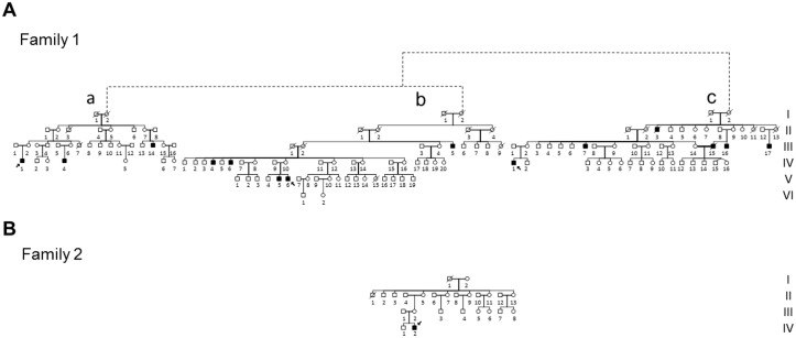
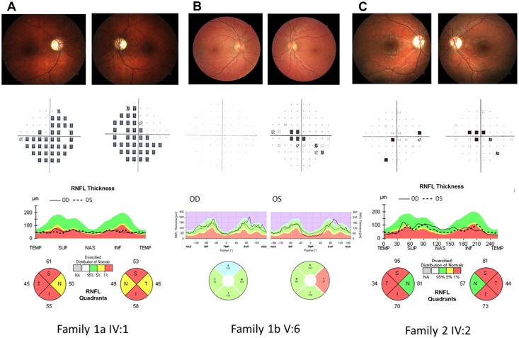
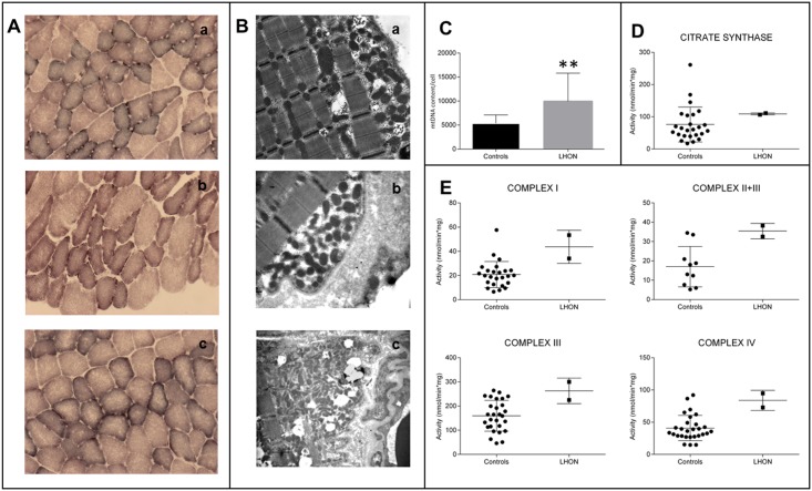
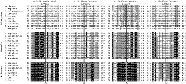
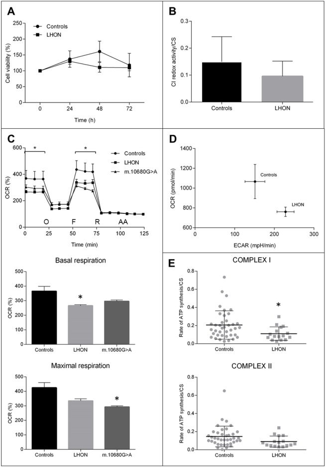
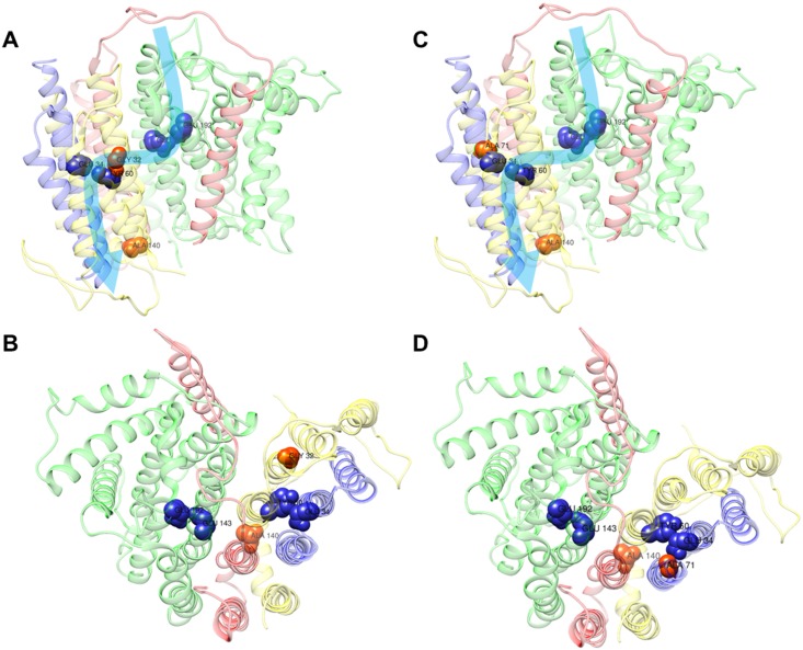
We here report on the existence of Leber's hereditary optic neuropathy (LHON) associated with peculiar combinations of individually non-pathogenic missense mitochondrial DNA (mtDNA) variants, affecting the MT-ND4, MT-ND4L and MT-ND6 subunit genes of Complex I. The pathogenic potential of these mtDNA haplotypes is supported by multiple evidences: first, the LHON phenotype is strictly inherited along the maternal line in one very large family; second, the combinations of mtDNA variants are unique to the two maternal lineages that are characterized by recurrence of LHON; third, the Complex I-dependent respiratory and oxidative phosphorylation defect is co-transferred from the proband's fibroblasts into the cybrid cell model. Finally, all but one of these missense mtDNA variants cluster along the same predicted fourth E-channel deputed to proton translocation within the transmembrane domain of Complex I, involving the ND1, ND4L and ND6 subunits. Hence, the definition of the pathogenic role of a specific mtDNA mutation becomes blurrier than ever and only an accurate evaluation of mitogenome sequence variation data from the general population, combined with functional analyses using the cybrid cell model, may lead to final validation. Our study conclusively shows that even in the absence of a clearly established LHON primary mutation, unprecedented combinations of missense mtDNA variants, individually known as polymorphisms, may lead to reduced OXPHOS efficiency sufficient to trigger LHON. In this context, we introduce a new diagnostic perspective that implies the complete sequence analysis of mitogenomes in LHON as mandatory gold standard diagnostic approach.


Conflict of interest statement

The authors have declared that no competing interests exist.

Figures

Fig 1
Fig 1. Pedigrees.
A. Pedigree of Family 1 with the reconstructed genealogy (indicated by dashed lines) of its three branches (a, b, c). Affected individuals are indicated in black; probands are indicated by arrows.B. Pedigree of Family 2. Affected individuals are indicated in black; proband is indicated by arrow.
Fig 2
Fig 2. Ophthalmological data.
A. Family 1a IV:1 Upper line: Fundus oculi pictures show bilateral optic nerve pallor. Middle line: Computerized visual fields (VFs) (pattern deviation) reveal bilateral generalized defect. Lower line: Optical coherence tomography (OCT) (Cirrus, Zeiss) demonstrates bilateral diffuse optic atrophy with relative nasal sparing. B. Family 1b V:6 Upper line: Fundus oculi pictures show normal optic nerve in OD and mild temporal pallor in OS. Middle line: Computerized VFs is unremarkable in OD and demonstrates a small central scotoma in OS. Lower line: OCT demonstrates normal retinal nerve fiber layer thickness in OD and temporal thinning in OS (Spectralis, Heidelberg). C. Family 2 IV:2 Upper line: Fundus oculi pictures show bilateral optic nerve pallor. Middle line: Computerized VFs reveal bilateral central scotoma. Lower line: OCT demonstrates bilateral diffuse optic atrophy with relative nasal sparing (Cirrus, Zeiss).
Fig 3
Fig 3. Morphological, molecular and biochemical analysis on skeletal muscle biopsies.
A. SDH staining of skeletal muscle biopsies from individuals IV:1 from Family 1a (a), V:6 from Family 1b (b) and IV:2 from Family 2 (c). B. Transmission electron microscopy of the same muscle specimens as in A. Both SDH histoenzymatic staining (A) and ultrastructural evaluation (B) demonstrate mitochondrial proliferation as highlighted respectively by subsarcolemmal increase of SDH reaction and corresponding accumulation of mitochondria. C. Assessment of mtDNA content per cell, presented as column with mean ± SD (n = 3; **p<0.001), confirms the activation of mitochondrial biogenesis as shown by the significant increase in LHON samples. D. The evaluation of citrate synthase activity, presented as scatter plot with mean ± SD, parallels again the results observed in C, with an increased mean value in the LHON samples. E. Evaluations of Complex I, Complex II+III, Complex III, Complex IV activities, presented as scatter plot with mean ± SD, reveal an increase of all activities in LHON samples.
Fig 4
Fig 4. Amino acid conservation analysis.
Global alignment of ND1, ND4L and ND6 protein sequences from a wide range of eukaryotes, mammals and primates. The neighborhoods (20 amino acids) of m.14582A>G/MT-ND6, m.14258G>A/MT-ND6, m.10680G>A/MT-ND4L and m.12033A>G/MT-ND4 are shown. Rectangles frame these specific variants. Amino acid residues with a percentage of conservation ranging between 70.0% and 79.9% are highlighted in light grey, those between 80.0% and 99.9% are highlighted in dark grey and those invariant (100%) are highlighted in black.
Fig 5
Fig 5. Biochemical characterization of cybrid clones.
A. Cell viability after different time of incubation in galactose medium (0, 24, 48, 72h). Data are expressed as percentage of T0 (n = 12; mean ± SEM). B. Rotenone sensitive redox activity of respiratory complex I normalized for CS activity (n = 9; mean ± SD). C. OCR traces as pmol O2/min, after the injection of 1μM oligomycin (O), 0.2μM FCCP (F), 1μM rotenone (R) and 1μM antimycin A (AA) (mean ± SEM). Asterisks indicate statistical significance (n> = 3; * p<0.05). D. XFe Metabolic Phenogram. Basal OCR (pmol/min) and ECAR (mpH/min) rates were plotted in controls vs LHON cybrids, showing a metabolic shift in LHON cybrids towards glycolysis. E. ATP synthesis rates normalized for CS activity driven by complex I substrates (malate/glutamate) and complex II substrate succinate (mean ± SD). Asterisks indicate statistical significance (n = 16; * p<0.05).
Fig 6
Fig 6. Complex I model.
Localization of polymorphic variants on the cryo-EM structure of the ovine complex I, [34] using the UCSF Chimera software. The ovine amino acids Ala140 (corresponding to human p.P139L, m.14258G>A/MT-ND6), Gly32 (corresponding to human p.V31A, m.14582A>G/MT-ND6) and Ala71 (corresponding to human p.A71T, m.10680G>A/MT-ND4L) are shown as red-labelled spheres; whereas residues Glu143/ND1, Glu192/ND1, Glu34/ND4L, Tyr60/ND6, the key residues for the E-channel (near Q site), are shown as blue-labelled spheres. The structures of ND1, ND4L, ND6 and ND3 subunits are shown as ribbons, in green, blue, yellow and red, respectively. The combination of variants in Family 1 (A-B) and Family 2 (C-D) are displayed as front (A-C) and upper (B-D) views. Light blue arrows indicate the proposed proton translocation pathway [34].

References

    1. Wallace DC, Singh G, Lott MT, Hodge JA, Schurr TG, Lezza AM, et al. Mitochondrial DNA mutation associated with Leber’s hereditary optic neuropathy. Science. 1988;242: 1427–1430. - PubMed
    1. Chinnery PF, Howell N, Andrews RM, Turnbull DM. Mitochondrial DNA analysis: polymorphisms and pathogenicity. J Med Genet. 1999;36: 505–510. - PMC - PubMed
    1. Montoya J, López-Gallardo E, Díez-Sánchez C, López-Pérez MJ, Ruiz-Pesini E. 20 years of human mtDNA pathologic point mutations: carefully reading the pathogenicity criteria. Biochim Biophys Acta. 2009;1787: 476–483. doi: 10.1016/j.bbabio.2008.09.003 - DOI - PubMed
    1. Carelli V, Ross-Cisneros FN, Sadun AA. Mitochondrial dysfunction as a cause of optic neuropathies. Prog Retin Eye Res. 2004;23: 53–89. doi: 10.1016/j.preteyeres.2003.10.003 - DOI - PubMed
    1. Yu-Wai-Man P, Griffiths PG, Chinnery PF. Mitochondrial optic neuropathies—disease mechanisms and therapeutic strategies. Prog Retin Eye Res. 2011;30: 81–114. doi: 10.1016/j.preteyeres.2010.11.002 - DOI - PMC - PubMed

Publication types

MeSH terms

Substances

LinkOut - more resources