. 2018 Apr 1;159(4):1570-1584.
doi: 10.1210/en.2018-00004.

GLP-1 Receptor Expression Within the Human Heart

Affiliations

GLP-1 Receptor Expression Within the Human Heart

Laurie L Baggio et al. Endocrinology. .

Abstract

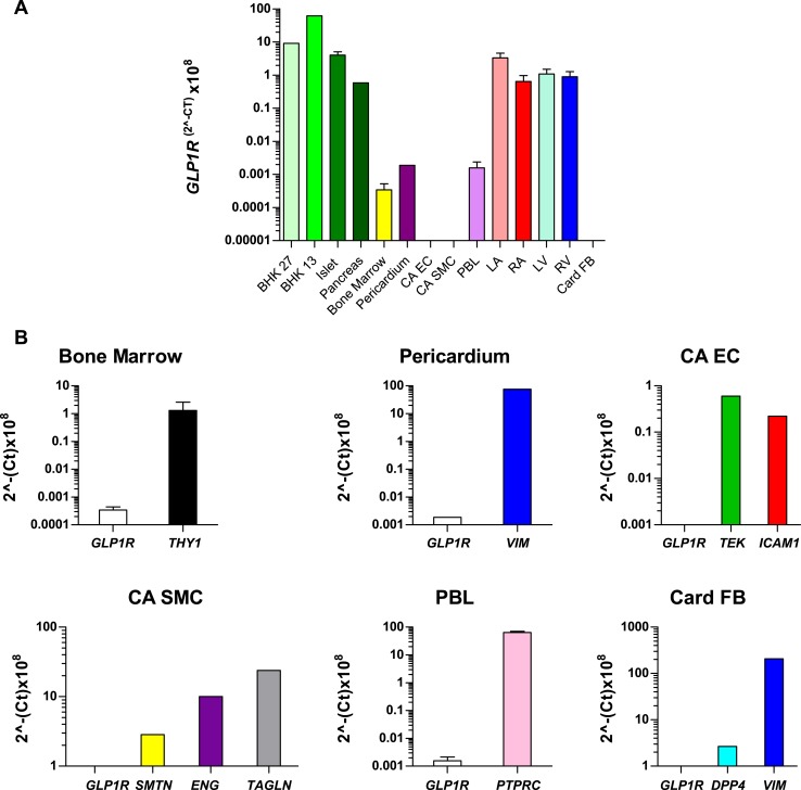
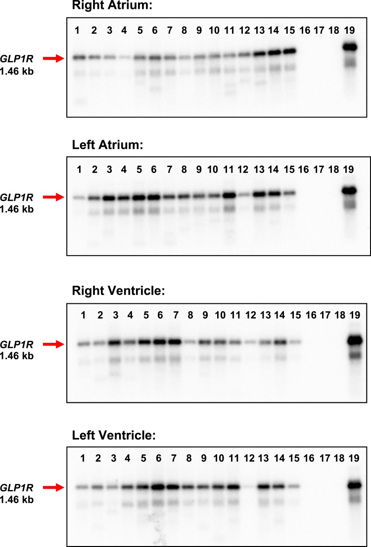
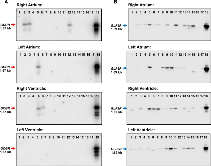
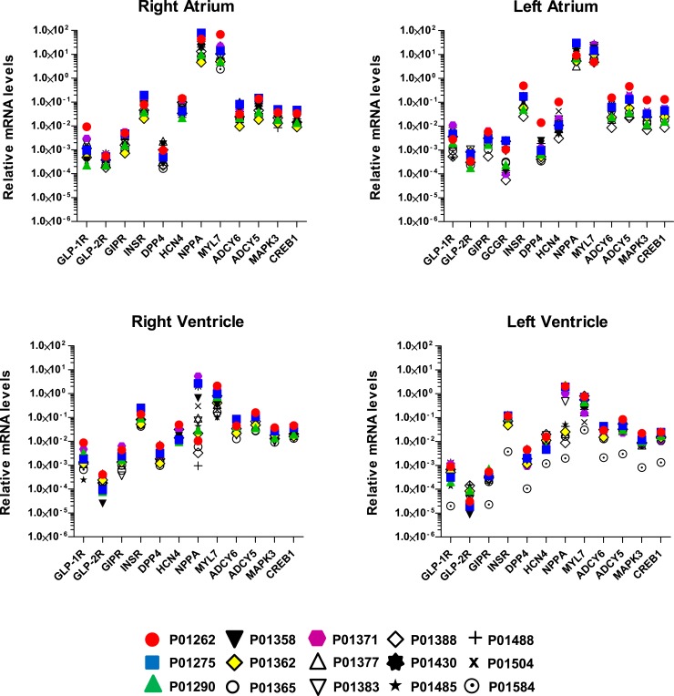
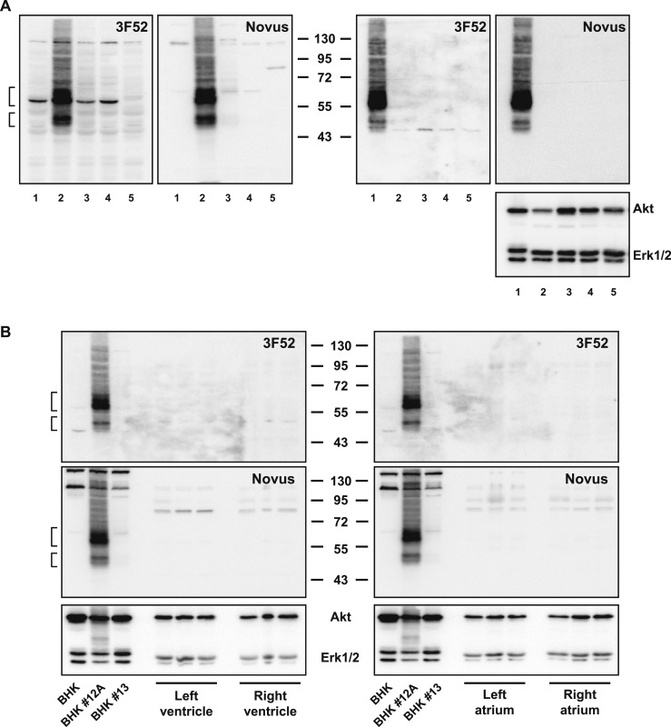
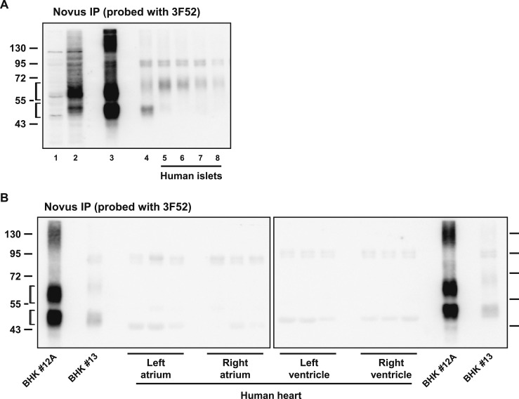
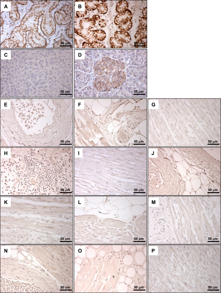
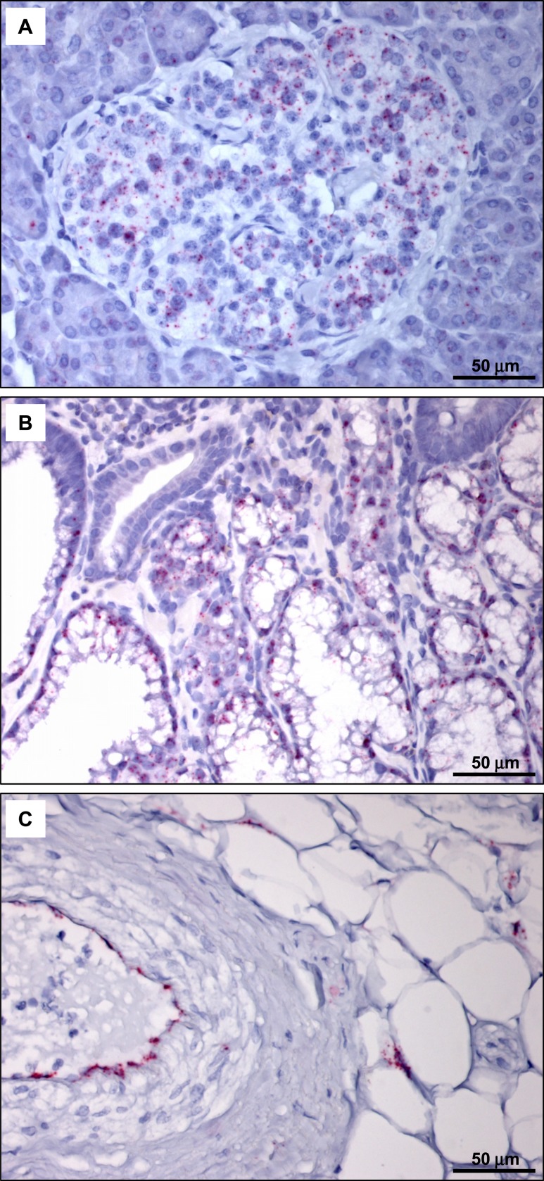
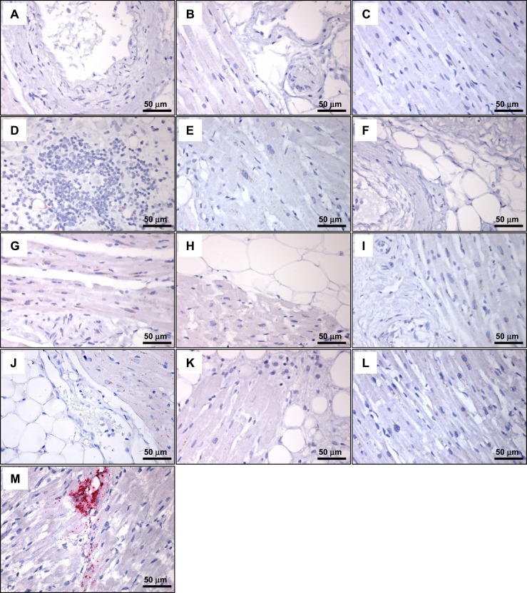
Glucagonlike peptide-1 receptor (GLP-1R) agonists, which are used to treat type 2 diabetes and obesity, reduce the rates of myocardial infarction and cardiovascular death. GLP-1R has been localized to the human sinoatrial node; however, its expression in ventricular tissue remains uncertain. Here we studied GLP-1R expression in the human heart using GLP-1R-directed antisera, quantitative polymerase chain reaction (PCR), reverse transcription PCR to detect full-length messenger RNA (mRNA) transcripts, and in situ hybridization (ISH). GLP1R mRNA transcripts, encompassing the entire open reading frame, were detected in all four cardiac chambers from 15 hearts at levels approximating those detected in human pancreas. In contrast, cardiac GLP2R expression was relatively lower, and cardiac GCGR expression was sporadic and not detected in the left ventricle. GLP1R mRNA transcripts were not detected in RNA from human cardiac fibroblasts, coronary artery endothelial, or vascular smooth muscle cells. Human Brunner glands and pancreatic islets exhibited GLP-1R immunopositivity and abundant expression of GLP1R mRNA transcripts by ISH. GLP1R transcripts were also detected by ISH in human cardiac sinoatrial node tissue. However, definitive cellular localization of GLP1R mRNA transcripts or immunoreactive GLP-1R protein within human cardiomyocytes or cardiac blood vessels remained elusive. Moreover, validated GLP-1R antisera lacked sufficient sensitivity to detect expression of the endogenous islet or cardiac GLP-1R by Western blotting. Hence, although human cardiac ventricles express the GLP1R, the identity of one or more ventricular cell type(s) that express a translated GLP1R protein requires further clarification with highly sensitive methods of detection.


Figures

Figure 1.
Figure 1.
GLP1R mRNA transcript levels in the human heart are comparable to those in human pancreas and islets. (A) GLP1R mRNA levels were measured via qPCR analysis in multiple human tissues and in transfected BHK cells that express low levels of the human GLP-1R. For data represented without standard error bars, each single RNA sample was analyzed in duplicate; for isolates depicted with error bars, peripheral blood lymphocyte samples were analyzed in duplicate from two different sources, and at least three different samples were analyzed in duplicate by qPCR for RNAs from islet, bone marrow, left atria (LA), right atria (RA), left ventricle (LV), and right ventricle (RV). (B) qPCR analysis of GLP1R and tissue- or cell-type–specific gene expression in the indicated samples as confirmation of RNA/cDNA integrity. For (A) and (B), data are expressed as cycle threshold (Ct) values because none of the housekeeping genes examined (ACTB, GPI, PSMB4, CHMP2A, and EMC7) exhibited consistent expression levels in all tissues examined. Values are mean ± standard error (where appropriate); n = 1 to 3 samples per tissue. LA samples are from patients P01371, P01430, and P01504. RA samples are from patients P01262, P01371, and P01377. LV samples are from patients P01262, P01430, and P01371. RV samples are from patients P01262, P01371, and P01504 (see Supplemental Table 1 and Figure 4). Islet samples are from donors R177, R199, and R200 (see Supplemental Table 3). CA EC, coronary artery endothelial cells; CA SMC, coronary artery smooth muscle cells; PBL, peripheral blood lymphocytes; Card FB, cardiac fibroblasts.
Figure 2.
Figure 2.
A full-length GLP1R is expressed in all four chambers of the human heart. RT-PCR analysis and Southern blotting using an internal GLP1R-specific oligonucleotide probe detected a 1.46-kb GLP1R mRNA transcript (red arrow) in the left and right atria and in the left and right ventricle of all 15 human hearts. Lanes 1 to 15, human heart samples; lane 16, negative control (H2O); lane 17, human cardiac fibroblasts; lane 18, human adipose tissue; lane 19, human pancreas (positive control). See Supplemental Table 1 for a description of the human heart samples.
Figure 3.
Figure 3.
GCGR and GLP2R mRNA expression in the human heart. RT-PCR analysis and Southern blotting using an internal oligonucleotide probe specific to each receptor RNA detected a 1.67-kb GCGR mRNA transcript (red arrow, left panels) in a small number of human hearts and a 1.69-kb GLP2R mRNA transcript (blue arrow, right panels) in all four cardiac chambers of several human heart samples. Lanes 1 to 15, human heart samples; lane 16, negative control (H2O); lane 17, human cardiac fibroblasts (GCGR blots) or human adipose tissue (GLP2R blots); lane 18 (positive control), human liver (GCGR blots) or human colon (GLP2R blots). See Supplemental Table 1 for a description of the human heart samples.
Figure 4.
Figure 4.
Quantitative gene expression profiles in all four chambers of the human heart. The mRNA levels of 12 or 13 different genes were measured in the four heart chambers from all 15 subjects via qPCR analysis. Data were normalized to β-actin. Each symbol represents analysis of RNA from an individual human heart (see Supplemental Table 1).
Figure 5.
Figure 5.
(A) Western blot analysis of whole cell or human islet tissue extracts using GLP-1R antibodies against the human GLP-1R, Mab 3F52, or polyclonal Novus. Molecular mass standards (kDa) are indicated in the center. Blots on the left contain whole cell extracts from BHK pcDNA3 (lane 1), BHK clone #12A (lane 2), BHK clone #13 (lane 3), BHK clone #27 (lane 4), and CFPAC-1 cells (lane 5). Blots on the right contain whole cell extracts from BHK clone #12A (lane 1) and whole tissue extracts from human islets #167 (lane 2), #177 (lane 3), #186 (lane 4), and 199 (lane 5). The blot on the far right was immunoblotted with Akt and Erk1/2 antibodies as loading controls. (B) Western blot analysis of whole cell or human cardiac tissue extracts analyzed using the indicated GLP-1R antibody. Molecular mass standards (kDa) are indicated in the center. Immunoblotting with Akt and Erk1/2 antibodies (bottom panels) was used as loading controls. In (A) and (B), the brackets indicate predicted migration positions of immunoreactive GLP-1R protein.
Figure 6.
Figure 6.
(A) Whole cell/tissue extracts after immunoprecipitation (IP) and Western blotting with the indicated GLP-1R antibodies. Lanes 1 and 2 are whole cell extracts, and lanes 3 through 8 correspond to immunoprecipitated samples. Lane 1, BHK pcDNA3; lane 2, BHK clone #12A; lane 3, BHK clone #12A; lane 4, BHK clone #13; lane 5, human islet #167; lane 6, human islet #177; lane 7, human islet #186; and lane 8, human islet #199. Molecular mass standards (kDa) are indicated on the left. (B) Whole tissue extracts from human heart chamber samples after IP and Western blotting with the indicated GLP-1R antibodies. Molecular mass standards (kDa) are indicated on the far left and right of the blots. In (A) and (B), the brackets indicate predicted migration positions of immunoreactive GLP-1R protein.
Figure 7.
Figure 7.
GLP-1R immunohistochemistry with Mab 3F52 does not detect GLP-1R–immunoreactive cells in human ventricular tissue. Immunostaining of hyperplastic human Brunner glands with isotype control antibody (A) and GLP-1R Mab 3F52 (B). The Novus 19400002 antibody did not reliably detect GLP-1R–immunopositive cells in sections containing human Brunner glands (data not shown). Immunostaining of human pancreas with isotype control antibody (C) and GLP-1R Mab 3F52 (D). (E–P) GLP-1R Mab 3F52 immunostaining of human cardiac tissues from individual subjects [(E) 193856; (F) 61704; (G) 70150; (H) 70847; (I) 81858; (J) 164011; (K) 116603; (L) 166469; (M) 179268; (N) 171263; (O) 182651; (P), 52411; see Supplemental Table 2].
Figure 8.
Figure 8.
ISH analysis detects GLP1R mRNA expression in human pancreas (A) and hyperplastic human Brunner glands (B). GLP1R-positive cells are indicated by punctate pink staining. (C) Detection of PECAM1 mRNA expression in human heart (case 164011; Supplemental Table 2) verifies the quality of the tissue and the integrity of the mRNA.
Figure 9.
Figure 9.
ISH analysis of GLP1R mRNA expression detects GLP1R-positive cells in human atrial tissue from the sinoatrial node region but not in human ventricular tissue. (A–L) ISH analysis of GLP1R mRNA expression in cardiac tissues from the following cases (see Supplemental Table 2): (A) 193856; (B) 61704; (C) 70150; (D) 70847; (E) 81858; (F) 164011; (G) 116603; (H) 166469; (I) 179268; (J) 171263; (K) 182651; (L) 52411. (M) ISH analysis of GLP1R mRNA expression in tissue from human sinoatrial node region.

Comment in

References

    1. Baggio LL, Drucker DJ. Biology of incretins: GLP-1 and GIP. Gastroenterology. 2007;132(6):2131–2157. - PubMed
    1. Campbell JE, Drucker DJ. Pharmacology, physiology, and mechanisms of incretin hormone action. Cell Metab. 2013;17(6):819–837. - PubMed
    1. Drucker DJ, Nauck MA. The incretin system: glucagon-like peptide-1 receptor agonists and dipeptidyl peptidase-4 inhibitors in type 2 diabetes. Lancet. 2006;368(9548):1696–1705. - PubMed
    1. Astrup A, Rössner S, Van Gaal L, Rissanen A, Niskanen L, Al Hakim M, Madsen J, Rasmussen MF, Lean ME; NN8022-1807 Study Group . Effects of liraglutide in the treatment of obesity: a randomised, double-blind, placebo-controlled study. Lancet. 2009;374(9701):1606–1616. - PubMed
    1. Davies M, Pieber TR, Hartoft-Nielsen ML, Hansen OKH, Jabbour S, Rosenstock J. Effect of oral semaglutide compared with placebo and subcutaneous semaglutide on glycemic control in patients with type 2 diabetes: a randomized clinical trial. JAMA. 2017;318(15):1460–1470. - PMC - PubMed

Publication types

MeSH terms

Substances

Grants and funding