. 2018 Feb 13;22(7):1810-1823.
doi: 10.1016/j.celrep.2018.01.058.

MicroRNA-223 Suppresses the Canonical NF-κB Pathway in Basal Keratinocytes to Dampen Neutrophilic Inflammation

Affiliations

MicroRNA-223 Suppresses the Canonical NF-κB Pathway in Basal Keratinocytes to Dampen Neutrophilic Inflammation

Wenqing Zhou et al. Cell Rep. .

Abstract

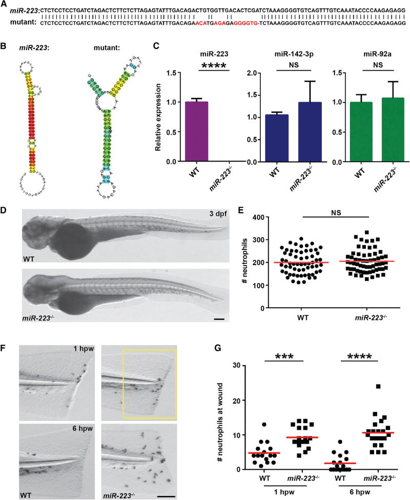
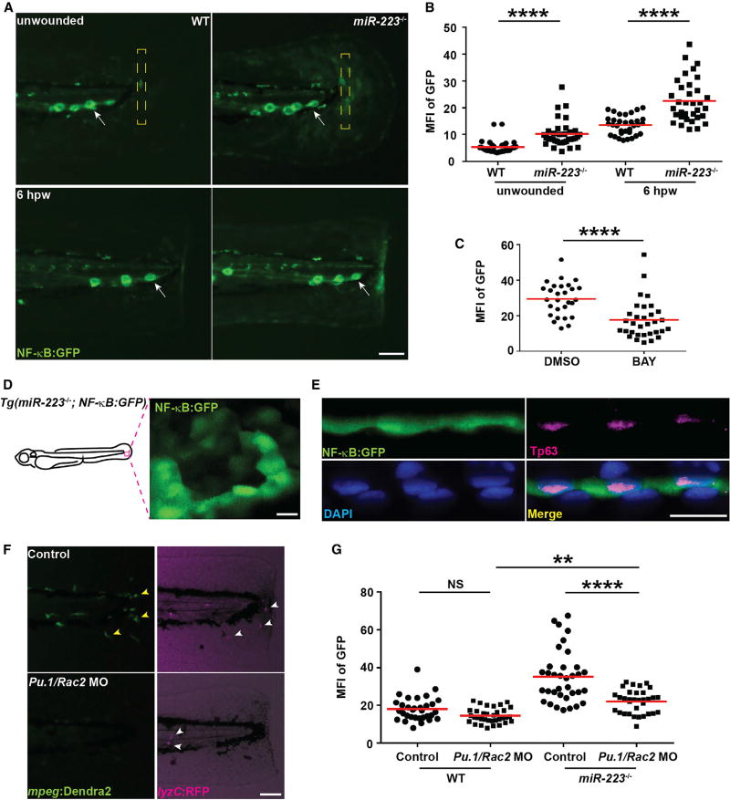
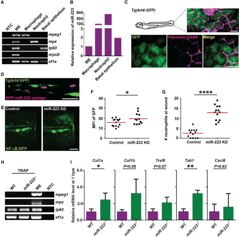
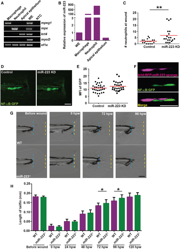
MicroRNA-223 is known as a myeloid-enriched anti-inflammatory microRNA that is dysregulated in numerous inflammatory conditions. Here, we report that neutrophilic inflammation (wound response) is augmented in miR-223-deficient zebrafish, due primarily to elevated activation of the canonical nuclear factor κB (NF-κB) pathway. NF-κB over-activation is restricted to the basal layer of the surface epithelium, although miR-223 is detected throughout the epithelium and in phagocytes. Not only phagocytes but also epithelial cells are involved in miR-223-mediated regulation of neutrophils' wound response and NF-κB activation. Cul1a/b, Traf6, and Tab1 are identified as direct targets of miR-223, and their levels rise in injured epithelium lacking miR-223. In addition, miR-223 is expressed in cultured human bronchial epithelial cells, where it also downregulates NF-κB signaling. Together, this direct connection between miR-223 and the canonical NF-κB pathway provides a mechanistic understanding of the multifaceted role of miR-223 and highlights the relevance of epithelial cells in dampening neutrophil activation.

Keywords: NF-κB; epithelium; inflammation; miR-223; microRNA; neutrophils; zebrafish.


Conflict of interest statement

DECLARATION OF INTERESTS

The authors declare no competing financial interests.

Figures

Figure 1
Figure 1. miR-223-Deficient Embryos Display Augmented Neutrophil Response to Tissue Injury
(A) Sequences of pre-mir-223 in WT and miR-223 mutant embryos. Mutated nucleotides are labeled in red. (B) The hairpin structures of miR-223 in WT and mutant embryos predicted by Centroidfold. (C) Expression of miR-223, miR-142-3p, and miR-92a in WT and miR-223−/− embryos determined by qRT-PCR. (D and E) Representative images (D) and quantification (E) of total neutrophil numbers in WT and miR-223−/− embryos. (F and G) Representative images (F) and quantification (G) of neutrophil recruitment to tail transection sites in WT and miR-223−/− embryos at 1 hpw and 6 hpw. The number of neutrophils in the boxed region were quantified. Scale bars, 100 µm. Data are representative of three independent experiments (E and G) or are pooled from three independent experiments (C). Mean ± SD. ***p < 0.001 and ****p < 0.0001, unpaired Student’s t test. See also Figures S1 and S2.
Figure 2
Figure 2. Neutrophil-Intrinsic miR-223 Regulates Neutrophilic Inflammation
(A) Schematics of Tol2-lyzC-Gal4-crys-CFP construct, injected into WT embryos to generate the transgenic line Tg(lyzC:Gal4-Vp16, crys:CFP)pu8; Tol2-UAS-miR-223 sponge that contains 6 bulged miR-223 binding sites after the UAS element and the Dendra2 control plasmid. (B and C) miR-223 sponge or Dendra2 control plasmids in (A) were injected into embryos from Tg(lyzC: Gal4-Vp16, crys:CFP)pu8 and Tg(lyzC: mCherry-H2B) cross. Tailfins were transected at 3 dpf. Representative images at indicating time points are shown in (B), and neutrophil recruitment at 6 hpw is shown in (C). (D) Schematics of Tol2-lyzC-miR-223/RFP constructs, injected into WT embryos to generate the transgenic line Tg(lyzC: RFP-miR-223)pu9 and the control line Tg(lyzC: RFP)pu10. (E) The transgenic lines illustrated in (D) were crossed into the miR-223−/− background. The siblings without RFP were used as negative control. miR-223 expression in indicated groups was quantified by qRT-PCR (mean ± SD). (F and G) Quantification of neutrophil recruitment to tail transection sites in embryos with miR-223 (F) or RFP control (G) expressed in neutrophils in the miR-223−/− background at 1 hpw and 6 hpw. Scale bars, 100 µm. One representative experiment of three independent repeats is shown. *p < 0.05, **p < 0.01, ***p < 0.001, and ****p < 0.0001, unpaired Student’s t test (C, F, and G) or one-way ANOVA (E). See also Movie S1.
Figure 3
Figure 3. miR-223 Regulates NF-κB Pathway by Suppressing Cul1a, Cul1b, Traf6, and Tab1
(A) Quantification of neutrophil recruitment at 6 hpw in miR-223−/− embryos injected with sgRNAs targeting Myd88, using gfp sgRNAs as the control. (B) Quantification of neutrophil recruitment at 6 hpw in miR-223−/− embryos treated with BAY (1 µM) or DMSO (1%). (C) Schematics of the canonical NF-κB signaling pathway. (D) The expression of Cul1a, Cul1b, Traf6, and Tab1 in WT and miR-223−/− embryos in unwounded embryos or at 1 hpw as determined by qRT-PCR. (E) Dual luciferase reporter assay showing specific suppression of Renilla luciferase depending on the 3′UTRs of Cul1a, Cul1b, Traf6, and Tab1 by miR-223. (F) Quantification of neutrophil recruitment at 6 hpw in miR-223−/− embryos injected with sgRNAs targeting both Cul1a and Cul1b or the gfp sgRNAs control. Data are representative of three independent experiments (A, B, and F) or are pooled from three independent experiments (D and E). Mean ± SD. *p < 0.05, **p < 0.01, and ***p < 0.001, unpaired Student’s t test and paired Student’s t test in (E). See also Figures S3–S5.
Figure 4
Figure 4. NF-κB Pathway Is Elevated in Basal Epithelial Cells in miR-223−/− Embryos
The NF-κB reporter line Tg(NF-κB:GFP) was crossed into the miR-223−/− and matched WT background. (A and B) Representative images (A) and quantification (B) of GFP signal. Mean fluorescence intensity (MFI) in the yellow square (A) in unwounded embryos or at the wound edge at 6 hpw was quantified. White arrows, neuromast cells constitutively expressing NF-κB signal. (C) Quantification of GFP signal at the wound edge at 6 hpw in miR-223−/− embryos treated with DMSO or BAY. (D) Representative confocal image of GFP+ cells in miR-223−/− embryos. (E) Immunofluorescence of GFP and Tp63 (basal cell marker) in Tg(miR-223−/−, NF-κB:GFP) embryos. Nucleus were stained with DAPI. Representative confocal images of vertical view are shown in (E). (F and G) Embryos from WT and miR-223−/− were injected with Pu.1 (200 µM) and Rac2 (100 µM) morpholinos. (F). Representative images showing the efficiency of the two morpholinos. Yellow arrowhead, macrophage; white arrowhead, neutrophils. (G) Quantification of GFP signal at the wound edge at 6 hpw in WT and miR-223−/− embryos. Scale bars, 100 µm (A and F), 20 µm (D and E). Data are representative of three independent experiments. **p < 0.01 and ****p < 0.0001, unpaired Student’s t test (B and C) or two-way ANOVA (G). See also Figure S6 and Movie S2.
Figure 5
Figure 5. miR-223 Suppresses NF-κB Activation in Basal Epithelial Cells in a Cell-Intrinsic Manner
(A and B) Macrophages, neutrophils, and basal epithelial cells were sorted from 3 dpf embryos. (A) RT-PCR of lineage-specific markers. mpeg, macrophage marker; mpx, neutrophil marker; tp63, basal epithelial cell marker; myoD, muscle cell marker; ef1a, loading control; WE, whole embryo; NTC, non-template control. (B) qRT-PCR of miR-223. (C) Tdtomato-CAAX mRNA was injected into Tg(krt4: GFP)pu11 embryos at 4-cell stage. Representative confocal images from the lateral view of 3 dpf larvae are shown. (D) RFP-miR-223 sponge mRNA was injected into Tg(krt4: GFP)pu11 embryos at 4-cell stage. A representative confocal image from the vertical view of 2 dpf larvae is shown. (E and F) Tg(NF-κB:GFP) were injected with miR-223 sponge or the RFP control mRNA at 4-cell stage. Representative images and quantification of GFP signal at the wound edge at 6 hpw are shown. (G) Quantification of neutrophil recruitment to the wound in embryos injected with miR-223 sponge or RFP mRNA at 4-cell stage. (H and I) Tol2-tp63-GFP-L10a was injected into WT or miR-223−/− embryos. At 3 dpf, cell-specific mRNA-ribosome complexes were isolated by anti-GFP antibodies at 1 hpw. (H) RT-PCR of lineage-specific markers as described in (A). (I) Real-time qPCR of indicated genes. Data are pooled from three independent experiments (mean + SD). Scale bars, 20 um (C and D), 100 µm (E). Data are representative of three independent experiments (F and G) or of two independent experiments (A and B). *p < 0.05, **p < 0.01, and ****p < 0.0001, unpaired Student’s t test.
Figure 6
Figure 6. Fin Regeneration Is Accelerated in miR-223-Deficient Embryos
(A) Apical epithelial cell were sorted from the Tg(krt4:GFP)pu11 and RT-PCR of lineage-specific markers were performed as described in Figure 5A. krt4, apical epithelial cell marker. (B) Real-time qPCR of miR-223. (C) Quantification of neutrophil recruitment at 6 hpw in embryos from Tg(krt4: RFP-miR-223 sponge)pu12 and Tg(krt4: RFP)pu13. (D and E) Tg(krt4: RFP-miR-223 sponge)pu12 and Tg(krt4: RFP)pu13 were crossed with Tg(NFB:GFP). Representative images and quantification of GFP signal at the wound edge at 6 hpw. (F) Representative confocal images of embryos from Tg(NFB:GFP/krt4: RFP-miR-223 sponge)pu12 at 6 hpw. (G and H) Tailfin regeneration in WT and miR-223−/− embryos. Tailfin transection was performed at 3 dpf. The length of regenerated tailfin was measured as the distance between the blue and yellow dash lines. (G) Representative images of tailfins at indicated time points. (H) Quantification of the length of tailfin at different time points (mean ± SD; n > 20 in each group). Scale bars, 100 µm (D and G), 20 um (F). Data are representative of three independent experiments (C, E, and H) or of two independent experiments (A and B). *p < 0.05, **p < 0.01, and ****p < 0.0001, unpaired Student’s t test.
Figure 7
Figure 7. miR-223 Regulates NF-κB Activation in Human Bronchial Epithelial Cells
(A) Real-time qPCR of miR-223 expression in human neutrophils, human immortalized bronchial epithelial cells (HBEC), transformed lung cancer epithelial cells (H441), and HEK293T. (B) Dual luciferase reporter assay showing specific suppression of Renilla luciferase dependent on 3′UTRs of human CUL1 and TAB2 by miR-223. (C) NF-κB activity in HEK293T cells expressing miR-223 or control with or without P. aeruginosa stimulation. NF-κB activity in HBEC cells expressing miR-223/control or miR-223-sponge/control with or without P. aeruginosa stimulation. Data are pooled from three independent experiments (mean ± SD). *p < 0.05, unpaired Student’s t test (A, C, and D) and paired Student’s t test (B).

References

    1. Aggad D, Stein C, Sieger D, Mazel M, Boudinot P, Herbomel P, Levraud JP, Lutfalla G, Leptin M. In vivo analysis of Ifn-γ1 and Ifn-γ2 signaling in zebrafish. J. Immunol. 2010;185:6774–6782. - PubMed
    1. Baek D, Villén J, Shin C, Camargo FD, Gygi SP, Bartel DP. The impact of microRNAs on protein output. Nature. 2008;455:64–71. - PMC - PubMed
    1. Bauernfeind F, Rieger A, Schildberg FA, Knolle PA, Schmid-Burgk JL, Hornung V. NLRP3 inflammasome activity is negatively controlled by miR-223. J. Immunol. 2012;189:4175–4181. - PubMed
    1. Borregaard N. Neutrophils, from marrow to microbes. Immunity. 2010;33:657–670. - PubMed
    1. Brantley DM, Chen CL, Muraoka RS, Bushdid PB, Bradberry JL, Kittrell F, Medina D, Matrisian LM, Kerr LD, Yull FE. Nuclear factor-kappaB (NF-kappaB) regulates proliferation and branching in mouse mammary epithelium. Mol. Biol. Cell. 2001;12:1445–1455. - PMC - PubMed

Publication types

MeSH terms