

hPMSC transplantation restoring ovarian function in premature ovarian failure mice is associated with change of Th17/Tc17 and Th17/Treg cell ratios through the PI3K/Akt signal pathway
- PMID: 29444704
- PMCID: PMC5813427
- DOI: 10.1186/s13287-018-0772-x
hPMSC transplantation restoring ovarian function in premature ovarian failure mice is associated with change of Th17/Tc17 and Th17/Treg cell ratios through the PI3K/Akt signal pathway
Retraction in
-
Retraction Note: hPMSC transplantation restoring ovarian function in premature ovarian failure mice is associated with change of Th17/Tc17 and Th17/Treg cell ratios through the PI3K/Akt signal pathway.Stem Cell Res Ther. 2022 Sep 14;13(1):471. doi: 10.1186/s13287-022-03173-8. Stem Cell Res Ther. 2022. PMID: 36104765 Free PMC article. No abstract available.
Abstract
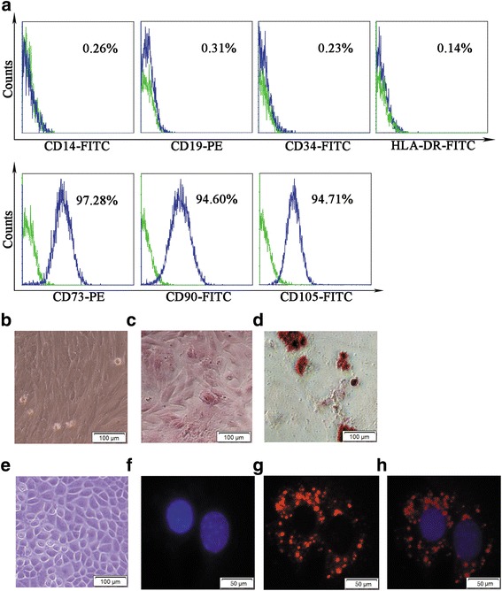
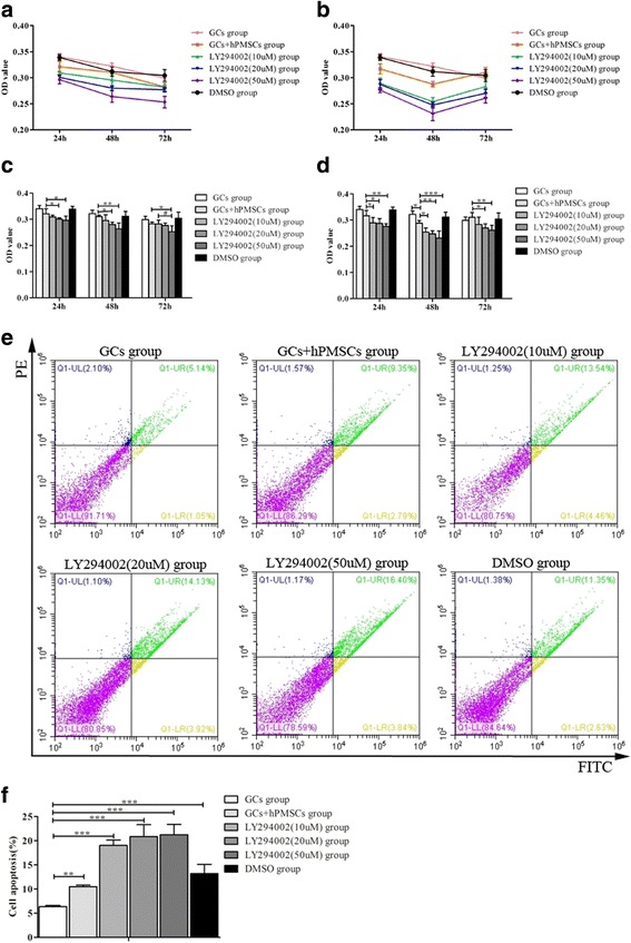
Background: Human placenta-derived mesenchymal stem cell (hPMSC) transplantation has been demonstrated to be an effective way of recovering ovarian function in mice with autoimmune induced premature ovarian failure (POF). But the exact mechanism remains unclear. The goal of the present study is to investigate the role of immune factors (T-helper 17 (Th17), cytotoxic T (Tc17) and regulatory T (Treg) cells) in the recovery of ovarian function and whether the phosphatidylinositol 3-kinase (PI3K)/Akt signal pathway is involved in the regulation.
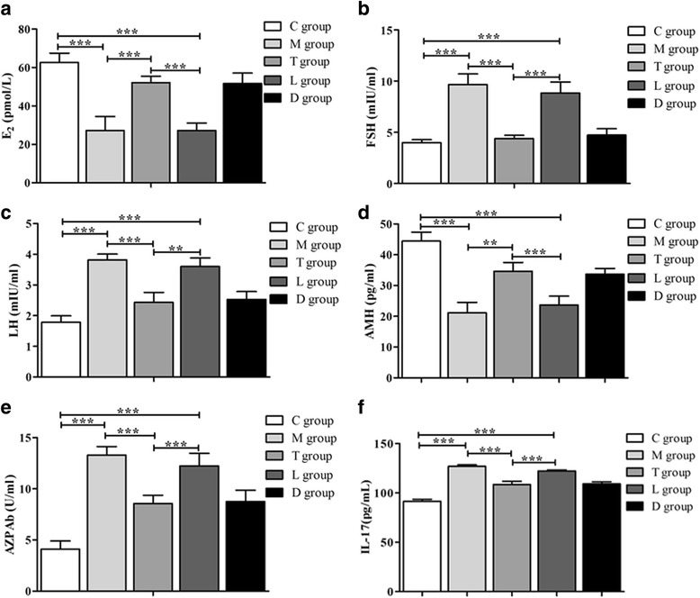
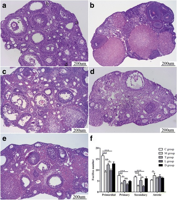
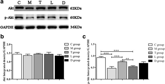
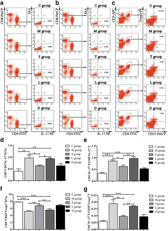
Methods: The inhibitor of PI3K/Akt was administered to observe its effect on ovarian function recovery and immune regulation. Serum levels of estradiol (E2), follicle stimulation hormone (FSH), luteinizing hormone (LH) and anti-Müllerian hormone (AMH)) and anti-Zona pellucida antibody (AZPAb) were measured by ELISA to evaluate ovarian function. The morphological changes of ovaries were observed by HE staining. Apoptosis of granular cells (GCs) was determined by detecting the expression of capase-3. Expression of p-Akt protein was detected by immunohistochemistry and western blot assay in ovarian tissues. The MTT assay was performed to assess GC proliferation. GC apoptosis was performed using flow cytometry analysis. Percentages of Th17, Tc17 and Treg cells were detected by flow cytometry. Expression of interleukin (IL)-17 in serum was measured by ELISA.
Results: LY294002 administration decreased serum levels of E2 and AMH, while the levels of FSH, LH and AZPAb in serum were increased compared with mice in the hPMSC transplantation group. The ovarian morphology presented as atrophy and fibrosis, with functional follicles exhausted. The expression of p-Akt in ovarian tissue was significantly decreased. Also, LY294002 administration significantly decreased proliferation and increased cell apoptosis in GCs, and for immune factors the ratios of Th17/Tc17 and Th17/Treg cells were significantly increased, as well as the serum levels of IL-17.
Conclusions: Our data suggest that the PI3K/Akt signal pathway is involved in the recovery of ovarian function by changing the ratios of Th17/ Tc17 and Th17/Treg cells in POF mice following hPMSC transplantation.
Keywords: Akt; Human placenta-derived mesenchymal stem cells; Immune factors; PI3K; Premature ovarian failure.
Conflict of interest statement
Authors’ information
No applicable.
Ethics approval and consent to participate
Animals were treated in accordance with the Basel Declaration in the context of phase experimental animals.
Consent for publication
Not applicable.
Competing interests
The authors declare that they have no competing interests.
Publisher’s Note
Springer Nature remains neutral with regard to jurisdictional claims in published maps and institutional affiliations.
Figures

References
-
- Elfayomy AK, Almasry SM, El-Tarhouny SA, Eldomiaty MA. Human umbilical cord blood-mesenchymal stem cells transplantation renovates the ovarian surface epithelium in a rat model of premature ovarian failure: possible direct and indirect effects. Tissue Cell. 2016;48:370–82. doi: 10.1016/j.tice.2016.05.001. - DOI - PubMed
-
- Muscari C, Bonafe F, Martin-Suarez S, Valgimigli S, Valente S, Fiumana E, Fiorelli F, Rubini G, Guarnieri C, Caldarera CM, Capitani O, Arpesella G, Pasquinelli G. Restored perfusion and reduced inflammation in the infarcted heart after grafting stem cells with a hyaluronan-based scaffold. J Cell Mol Med. 2013;17:518–30. doi: 10.1111/jcmm.12039. - DOI - PMC - PubMed
-
- Luan X, Li G, Wang G, Wang F, Lin Y. Human placenta-derived mesenchymal stem cells suppress t cell proliferation and support the culture expansion of cord blood CD34(+) cells: a comparison with human bone marrow-derived mesenchymal stem cells. Tissue Cell. 2013;45:32–8. doi: 10.1016/j.tice.2012.09.002. - DOI - PubMed
-
- Yin N, Zhao W, Luo Q, Yuan W, Luan X, Zhang H. Restoring ovarian function with human placenta-derived mesenchymal stem cells in autoimmune-induced premature ovarian failure mice mediated by Treg cells and associated cytokines. Reprod Sci (Thousand Oaks, Calif) 2017:1933719117732156. 10.1177/1933719117732156. [Epub ahead of print]. - PubMed
Publication types
MeSH terms
Substances
LinkOut - more resources
Full Text Sources
Other Literature Sources
Medical
Miscellaneous
