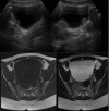
Isolated bladder schwannoma: a rare presentation
- PMID: 29444793
- PMCID: PMC5847897
- DOI: 10.1136/bcr-2017-223154
Isolated bladder schwannoma: a rare presentation
Abstract
Bladder schwannoma is a rare tumour arising from Schwann cells in nerve sheaths. It is usually more common in patients diagnosed with neurofibromatosis. However, isolated cases of urinary bladder schwannoma is incredibly rare, attributing to <0.1% of bladder tumours. A literature review and analysis revealed that it presents in adulthood, is mostly symptomatic and diagnosis is established histologically. We report a case of isolated bladder schwannoma in 25 year-old female who presented with dyspareunia.
Keywords: peripheral nerve disease; urinary and genital tract disorders; urological surgery; urology.
© BMJ Publishing Group Ltd (unless otherwise stated in the text of the article) 2018. All rights reserved. No commercial use is permitted unless otherwise expressly granted.
Conflict of interest statement
Competing interests: None declared.
Figures


References
-
- Pytel P, Anthony DC. Peripheral nerves and skeletal muscles Robbins and Cotran pathologic basis of disease. 9th ed Philadelphia: Elsevier Saunders, 2015:1246–9.
Publication types
MeSH terms
LinkOut - more resources
Full Text Sources
Other Literature Sources
Medical
Research Materials
