Review
. 2018 Feb;8(2):170216.
doi: 10.1098/rsob.170216.

Rett syndrome: a neurological disorder with metabolic components

Affiliations
Review

Rett syndrome: a neurological disorder with metabolic components

Stephanie M Kyle et al. Open Biol. 2018 Feb.

Abstract

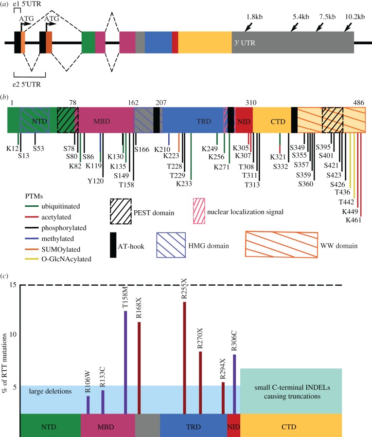
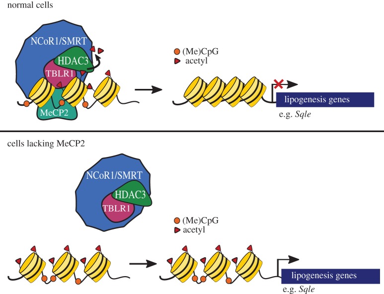
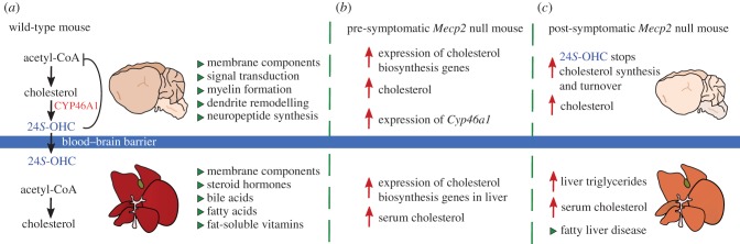
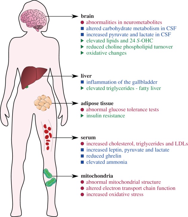
Rett syndrome (RTT) is a neurological disorder caused by mutations in the X-linked gene methyl-CpG-binding protein 2 (MECP2), a ubiquitously expressed transcriptional regulator. Despite remarkable scientific progress since its discovery, the mechanism by which MECP2 mutations cause RTT symptoms is largely unknown. Consequently, treatment options for patients are currently limited and centred on symptom relief. Thought to be an entirely neurological disorder, RTT research has focused on the role of MECP2 in the central nervous system. However, the variety of phenotypes identified in Mecp2 mutant mouse models and RTT patients implicate important roles for MeCP2 in peripheral systems. Here, we review the history of RTT, highlighting breakthroughs in the field that have led us to present day. We explore the current evidence supporting metabolic dysfunction as a component of RTT, presenting recent studies that have revealed perturbed lipid metabolism in the brain and peripheral tissues of mouse models and patients. Such findings may have an impact on the quality of life of RTT patients as both dietary and drug intervention can alter lipid metabolism. Ultimately, we conclude that a thorough knowledge of MeCP2's varied functional targets in the brain and body will be required to treat this complex syndrome.

Keywords: Rett syndrome; histone deacetylase; metabolism; methyl-CpG-binding protein 2; nuclear corepressor.


Conflict of interest statement

M.J.J. is a scientific founder of ArRETT Neurosciences, Inc., a company designed to provide treatments to Rett syndrome patients.

Figures

Figure 1.
Figure 1.
Timeline of stages and symptom onset in RTT patients. Rett syndrome (RTT) is divided into four progressive stages. Patients display seemingly normal early development. Between 6 and 18 months of age, patients experience a period of developmental stagnation (Stage I) and no longer meet their mental, cognitive or motor milestones. Head circumference growth slows and this period lasts for weeks to months. Stage II is defined by rapid developmental regression in which acquired purposeful hand movements and verbal skills are lost. Microcephaly worsens and breathing irregularities and/or seizures arise. Stage III is a pseudo-stationary plateau period in which patients may show mild recovery in cognitive interests, but purposeful hand and body movements remain severely diminished. Stage IV is defined by motor deterioration and may last decades. Many patients are wheelchair and/or gastrostomy-tube dependent. However, not all girls progress to this severe stage.
Figure 2.
Figure 2.
Mutations in the multifunctional protein MeCP2 cause RTT. Coloured boxes indicate different encoded functional domains: light orange, N-terminus of MeCP2_e1; dark orange, N-terminus of MeCP2_e2; green, N-terminal domain (NTD) which has identical amino acid sequences between the two isoforms; pink, methyl-binding domain (MBD); blue, transcriptional repression domain (TRD); red, nuclear coreceptor co-repressor (NCoR) interaction domain (NID); yellow, C-terminal domain (CTD). (a) The four exons in the MECP2 gene. Arrows in exons 1 and 2 indicate the ‘ATG’ start codons for MeCP2_e1 or MeCP2_e2, respectively. Arrows in the 3′ UTR indicate multiple polyA sites resulting in different-length transcripts. Dashed lines on the top indicate the splicing pattern of MeCP2_e1 and dashed lines on the bottom indicate the splicing pattern of MeCP2_e2. (b) Functional domains and post-translational modifications (PTMs) of MeCP2. Coordinates are in relation to isoform MeCP2_e2. MeCP2 contains two PEST domains (black slashed boxes), two HMG domains (blue slashed boxes), three AT-hook domains (black solid boxes), one functional nuclear localization signal (NLS) (pink slashed box) and one WW domain (orange slashed box). PTMs are scattered throughout the protein and regulate interactions with MeCP2 binding partners. (c) Common damaging MECP2 mutations. Schematic of MeCP2 with functional domains. y-axis represents percentage of RTT patients with indicated mutation. Missense mutations are in purple and nonsense mutations are in red. Combined, these point mutations make up approximately 70% of all RTT-causing mutations.
Figure 3.
Figure 3.
MeCP2 anchors the NCoR/SMRT to methylated DNA. In healthy cells, MeCP2 binds methylated CpG dinucleotides (orange circles) and recruits the NCoR1/SMRT-HDAC3 co-repressor complex to regulatory sites surrounding the target loci. HDAC3 removes acetylation marks from surrounding histones to compact chromatin and prevent transcription of target genes. In Mecp2 mutant cells, the NCOR1/SMRT-HDAC3 complex cannot bind to methylated DNA resulting in an open chromatin state and increased transcription of genes. Known target genes of the complex in the liver include Sqle and other lipogenesis enzymes. Targets in the brain remain unknown.
Figure 4.
Figure 4.
Cholesterol metabolism is perturbed in Mecp2 mutant mice. (a) In the wild-type mouse, the brain produces cholesterol in situ as cholesterol cannot pass the blood–brain barrier (BBB). Acetyl-CoA enters the cholesterol biosynthesis pathway to make cholesterol which has many essential functions (green triangles). The enzyme CYP46A1 converts excess cholesterol into 24S-hydroxycholesterol (24S-OHC) for one-way egress across the BBB. The liver participates in cholesterol biosynthesis to provide cholesterol to other tissues in the body. (b) In pre-symptomatic Mecp2 null mice (3–4 weeks old), increased expression of cholesterol biosynthesis genes in the brain leads to elevated brain cholesterol levels. Consequently, the expression of Cyp46a1 is increased, indicating a heightened need for cholesterol turnover. Outside of the central nervous system, serum cholesterol is elevated, and expression of cholesterol biosynthesis genes is elevated in the liver. (c) In symptomatic Mecp2 null mice (8–10 weeks old), the brain is smaller due to lack of Mecp2. Cholesterol biosynthesis decreases drastically in the brain, likely due to feedback from elevated 24S-OHC. Owing to decreased synthesis, brain cholesterol remains high, but not as high as at younger ages (smaller red arrow). Serum cholesterol and/or triglycerides may also be elevated, depending upon genetic background. Triglycerides accumulate in the liver, and fatty liver disease develops, as indicated by pale liver.
Figure 5.
Figure 5.
Metabolic components of Rett syndrome. Common metabolic disturbances observed in RTT patients (blue squares), Mecp2-mutant mouse models (green triangles) that may be a feature of human disease, or in both human and mouse (pink circles). Note that metabolic parameters may vary within the patient population or among different mouse strains: for example, hyperammonaemia was found in a subset of patients, but was dropped as a diagnostic criterion because it was not common. Such findings suggest that genetic variation may play a big role in the penetrance of all but key diagnostic features.

References

    1. Rett A. 1966. On a unusual brain atrophy syndrome in hyperammonemia in childhood. Wien. Med. Wochenschr. 1946, 723–726. - PubMed
    1. Hagberg B, Aicardi J, Dias K, Ramos O. 1983. A progressive syndrome of autism, dementia, ataxia, and loss of purposeful hand use in girls: Rett's syndrome: report of 35 cases. Ann. Neurol. 14, 471–479. (doi:10.1002/ana.410140412) - DOI - PubMed
    1. Burd L, Randall T, Martsolf JT, Kerbeshian J. 1991. Rett syndrome symptomatology of institutionalized adults with mental retardation: comparison of males and females. Am. J. Ment. Retard. 95, 596–601. - PubMed
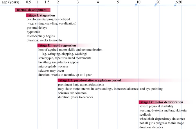
    1. Hagberg B. 2002. Clinical manifestations and stages of Rett syndrome. Ment. Retard. Dev. Disabil. Res. Rev. 8, 61–65. (doi:10.1002/mrdd.10020) - DOI - PubMed
    1. Hagberg B, Witt-Engerström I, Opitz JM, Reynolds JF. 1986. Rett syndrome: a suggested staging system for describing impairment profile with increasing age towards adolescence. Am. J. Med. Genet. 25, 47–59. (doi:10.1002/ajmg.1320250506) - DOI - PubMed

LinkOut - more resources