Review
. 2018 Feb 14;9(2):228.
doi: 10.1038/s41419-018-0313-7.

The carnitine system and cancer metabolic plasticity

Affiliations
Review

The carnitine system and cancer metabolic plasticity

Mariarosa Anna Beatrice Melone et al. Cell Death Dis. .

Abstract

Metabolic flexibility describes the ability of cells to respond or adapt its metabolism to support and enable rapid proliferation, continuous growth, and survival in hostile conditions. This dynamic character of the cellular metabolic network appears enhanced in cancer cells, in order to increase the adaptive phenotype and to maintain both viability and uncontrolled proliferation. Cancer cells can reprogram their metabolism to satisfy the energy as well as the biosynthetic intermediate request and to preserve their integrity from the harsh and hypoxic environment. Although several studies now recognize these reprogrammed activities as hallmarks of cancer, it remains unclear which are the pathways involved in regulating metabolic plasticity. Recent findings have suggested that carnitine system (CS) could be considered as a gridlock to finely trigger the metabolic flexibility of cancer cells. Indeed, the components of this system are involved in the bi-directional transport of acyl moieties from cytosol to mitochondria and vice versa, thus playing a fundamental role in tuning the switch between the glucose and fatty acid metabolism. Therefore, the CS regulation, at both enzymatic and epigenetic levels, plays a pivotal role in tumors, suggesting new druggable pathways for prevention and treatment of human cancer.


Conflict of interest statement

The authors declare that they have no conflict of interest.

Figures

Fig. 1
Fig. 1. Schematic view of carnitine system.
The long-chain fatty acid-CoAs are converted into the carnitine derivatives by CPT1, located on the outer mitochondrial membrane of the contact sites. A specific carnitine-acylcarnitine translocase (CACT) catalyzes the mole-to-mole exchange of carnitine/acetylcarnitine and acylcarnitines promoting the import of the acylcarnitines through the mitochondrial membranes. In the mitochondrial matrix, long-chain acylcarnitines are reconverted to the respective long-chain acyl-CoAs by carnitine-palmitoyltransferase-2 (CPT2) and undergo β-oxidation to produce acetyl-CoAs. Finally, CrAT converts short-chain acetyl-CoAs to their membrane permeant acetylcarnitine counterparts, allowing CACT to export them from mitochondrion to cytoplasm
Fig. 2
Fig. 2. Epigenetic ACC2 control modulates the reprogramming of fatty acid metabolism in cancer cells.
a Epigenetic control of ACACB by histone acetylation induces an increased expression of both ACC2 and its catalytic product, malonyl-CoA, leading to the inhibition of CPT1A activity. On the contrary, b histone deacetylation by sirtuin(s) (SIRT1/6), also promoted by lactate-induced acidification of the microenvironment, leads to a decreased expression of ACACB and consequently enhances CPT1A activity. NADH and acetyl-CoA, two metabolic intermediates produced in the course of FAO, promote activation of PDK, which in turn phospho-inactivates the E1α subunit of the PDH complex, leading to lower rates of glucose oxidation and higher rates of lactate release
Fig. 3
Fig. 3. The involvement of carnitine cycle in cancer cell metabolism.
In cancer metabolism the aerobic glycolysis induces the conversion of the pyruvate (the end product of the glycolysis) into lactate (shown in orange) leading to the acidification of the microenvironment. The increased acidosis (shown in blue), in association with epigenetic mechanism(s), promotes the downregulation of the mitochondrial ACC2 isoform that in turn increases FAO (shown in red) via CPT1A upregulation. NADH and acetyl-CoA, produced in excess during FAO, promote the conversion of pyruvate into lactate through the inhibition of PDH by PDK (Randle effect). The increased availability of mitochondrial acetyl-CoA enhances the intra-mitochondrial non-enzymatic acetylation of proteins both of the Kreb’s cycle and of the carnitine cycle, avoiding the risk associated with mitochondria overfeeding. In addition, the excess of acetyl-CoA is exported in the cytosol either as citrate or as acetyl l-carnitine (ALCAR). The citrate is converted into acetyl-CoA by ACLY for the synthesis of fatty acids that might be re-imported into the mitochondria for beta-oxidation (Futile Cycle). ALCAR, shuttled to the nucleus (shown in violet), can be used as source of acetyl groups for histone acetylation, probably contributing to lipid metabolism-specific gene expression
Fig. 4
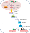
Fig. 4. miRNAs biogenesis and mechanism of action.
MicroRNAs are transcribed by RNA polymerases II and III in pri-miRNAs, generating precursors that undergo a series of cleavage events to form mature microRNA. Drosha, the first nuclear ribonuclease III, recognizes pri-miRNA and cuts the double-stranded RNA freeing a pre-miRNA. Pre-miRNA hairpin is exported from the nucleus in a process involving the nucleocytoplasmic protein Exportin-5. In the cytoplasm, the pre-miRNA hairpin is cleaved by the RNase III enzyme Dicer in an miRNA duplex of 18–22 nt. Although either the duplex strands may potentially act as functional miRNAs, only one strand is usually incorporated into the RNA-induced silencing complex (RISC) where the miRNA and its mRNA target interact. Mature miRNA acts either by degrading the mRNA target or by inhibiting its translation
Fig. 5
Fig. 5. MiRNAs influence the carnitine system components.
MiRNAs regulate cell metabolic plasticity by modulating the expression of enzymes involved in several metabolic pathways. MiRNAs affect both directly and indirectly the carnitine system components. Mir-33a/b and miR-122 target AMPK (activated by metabolic stress) and ACC1/2 respectively, whereas miR-205 targets the acyl-CoA synthetase, indirectly regulating the components of carnitine system. In addition, the carnitine system components are directly regulated by miR-370, miR-124 (CPT1A), miR-129 (CACT), miR-33a/b (CPT1A and CrAT), and miR-378 (CrAT)

References

    1. Goodpaster BH, Sparks LM. Metabolic flexibility in health and disease. Cell Metab. 2017;25:1027–1036. doi: 10.1016/j.cmet.2017.04.015. - DOI - PMC - PubMed
    1. Quail D, Joyce J. Microenvironmental regulation of tumor progression and metastasis. Nat. Med. 2013;19:1423–1437. doi: 10.1038/nm.3394. - DOI - PMC - PubMed
    1. Pavlova NN, Thompson CB. The emerging hallmarks of cancer metabolism. Cell Metab. 2016;23:27–47. doi: 10.1016/j.cmet.2015.12.006. - DOI - PMC - PubMed
    1. Martinez-Reyes I, et al. TCA cycle and mitochondrial membrane potential are necessary for diverse biological functions. Mol. Cell. 2016;61:199–209. doi: 10.1016/j.molcel.2015.12.002. - DOI - PMC - PubMed
    1. Gao X, Reid MA, Kong M, Locasale JW. Metabolic interactions with cancer epigenetics. Mol. Asp. Med. 2016;54:50–57. doi: 10.1016/j.mam.2016.09.001. - DOI - PMC - PubMed

Publication types

MeSH terms