. 2018 Feb 14;8(1):3019.
doi: 10.1038/s41598-018-21329-6.

Kinesin 1 regulates cilia length through an interaction with the Bardet-Biedl syndrome related protein CCDC28B

Affiliations

Kinesin 1 regulates cilia length through an interaction with the Bardet-Biedl syndrome related protein CCDC28B

Rossina Novas et al. Sci Rep. .

Abstract

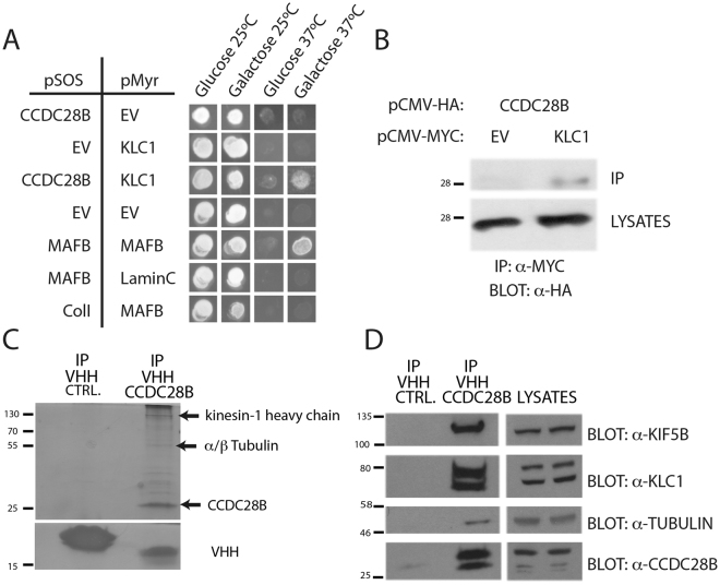
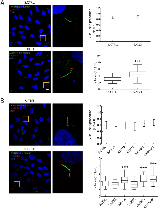
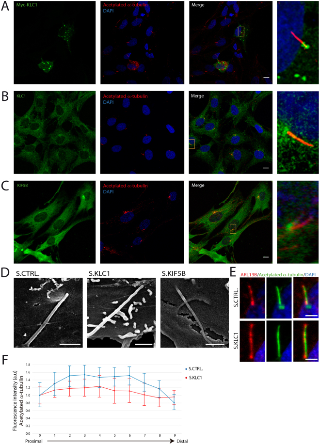
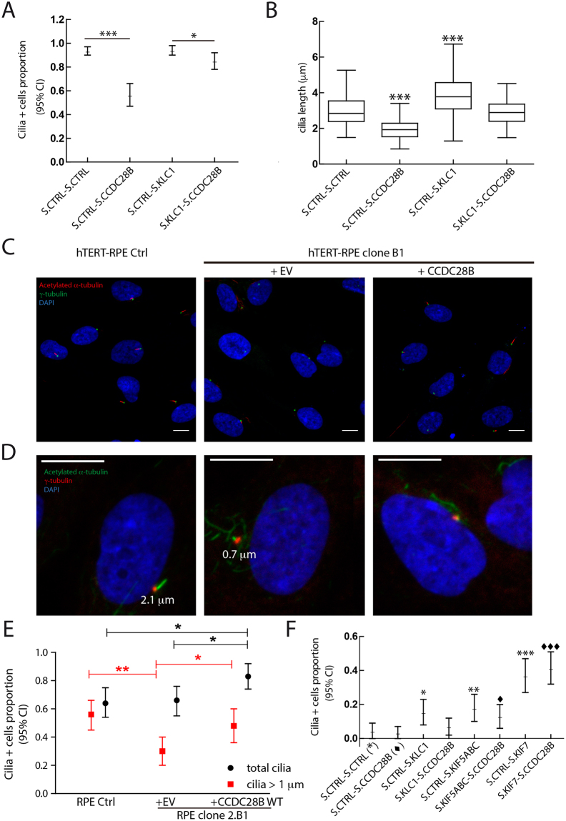
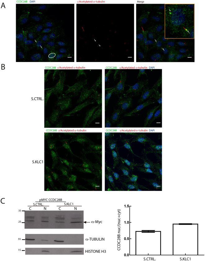
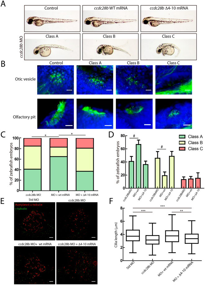
Bardet-Biedl syndrome (BBS) is a ciliopathy characterized by retinal degeneration, obesity, polydactyly, renal disease and mental retardation. CCDC28B is a BBS-associated protein that we have previously shown plays a role in cilia length regulation whereby its depletion results in shortened cilia both in cells and Danio rerio (zebrafish). At least part of that role is achieved by its interaction with the mTORC2 component SIN1, but the mechanistic details of this interaction and/or additional functions that CCDC28B might play in the context of cilia remain poorly understood. Here we uncover a novel interaction between CCDC28B and the kinesin 1 molecular motor that is relevant to cilia. CCDC28B interacts with kinesin light chain 1 (KLC1) and the heavy chain KIF5B. Notably, depletion of these kinesin 1 components results in abnormally elongated cilia. Furthermore, through genetic interaction studies we demonstrate that kinesin 1 regulates ciliogenesis through CCDC28B. We show that kinesin 1 regulates the subcellular distribution of CCDC28B, unexpectedly, inhibiting its nuclear accumulation, and a ccdc28b mutant missing a nuclear localization motif fails to rescue the phenotype in zebrafish morphant embryos. Therefore, we uncover a previously unknown role of kinesin 1 in cilia length regulation that relies on the BBS related protein CCDC28B.


Conflict of interest statement

The authors declare no competing interests.

Figures

Figure 1
Figure 1
CCDC28B interacts with the kinesin 1 components KLC1 and KIF5B. (A) The co-expression of CCDC28B and KLC1 allows cdc25H yeast cells to grow at the non-permissive temperature of 37 °C only on galactose that triggers expression from the pMyr construct. Controls: MAFB-MAFB (positive); MAFB-Lamin C (negative), ColI-MAFB (negative), pSOS EV-pMyr EV (negative) and pSOS CCDC28B-pMyr EV (negative). (B) HA-CCDC28B is detected only in the Myc-KLC1 immunoprecipitate. Cell lysates are shown to control for protein input. Bands were cropped from the same blot which is shown in Fig. S2A. (C) Immunoprecipitation of overexpressed CCDC28B in Hek293 cells with the specific single domain llama antibody (VHH) results in the co-immunoprecipitation of additional proteins. Arrows indicate gel bands analyzed by mass spectrometry. An irrelevant VHH was used as control. The gel was cut in two to avoid saturation of the VHH bands when silver-staining the upper part of the gel. (D) The VHH against CCDC28B was used for immunoprecipitation and specific antibodies were used to detect KIF5B, KLC1, α-tubulin and CCDC28B. Cell lysates show the corresponding proteins in the extracts used for immunoprecipitation. Bands shown were cropped from the original blot. The full-length membrane was cut and exposed to the different antibodies (see Fig. S2B,C for blots and for details).
Figure 2
Figure 2
Depletion of KLC1 and KIF5B results in elongated cilia. hTERT-RPE cells were analyzed by confocal microscopy using anti-acetylated tubulin (green), anti γ-tubulin (red) and DAPI (blue) to stain cilia, basal bodies and nuclei respectively. Cilia length was measured and results are expressed as box plots. Results are representative of three independent experiments. (A) While knockdown of KLC1 did not affect the proportion of cilia-positive cells compared to a control stealth (hypothesis test for proportions), it did result in significantly elongated cilia (statistical test: Mann-Whitney; ***P < 0.0001). At least 100 cilia were measured per condition (143 for S.Ctrl. and 147 for S.KLC1). (B) Knockdown of the different KIF5s (S.KIF5A, B, C) did not affect the proportion of cilia-positive cells. Similarly to KLC1 KD, depletion of KIF5B, KIF5BC and KIF5ABC resulted in elongated cilia (122 KIF5B KD, 96 KIF5BC and 109 KIF5ABC cilia were measured and compared to 95 control). Statistical test: one-way ANOVA. Asterisks denote statistical significant differences compared to controls. ***P < 0.0001. Scale bars correspond to 10 μm.
Figure 3
Figure 3
KLC1 and KIF5B are found at the base of cilia. Confocal microscopy analysis of hTERT-RPE cells. (A) Cells were transfected to overexpress Myc-tagged KLC1. In addition to cytoplasmic aggregates, a pool of Myc-KLC1 (anti-Myc, green) is found at the base of cilia (anti acetylated α-tubulin, red). (B) In addition to a diffuse cytoplasmic signal, the anti-KLC1 antibody shows an accumulation of endogenous protein at the base of cilia. (C) Similarly, endogenous KIF5B is found in the cytoplasm and also concentrated at the base of cilia. Yellow boxes mark the area that is magnified and showed in panels on the right. DAPI was used to stain nuclei. Scale bars correspond to 10 μm. (D) Scanning electron micrographs showing cilia in hTERT-RPE cells transfected with control (S.CTRL), KLC1 (S.KLC1) or KIF5B (S.KIF5B) stealth RNA oligos. Scale bars correspond to 1 μm. (E,F) The level of tubulin acetylation in control and KLC1 KD cilia was quantified measuring the fluorescence intensity of the signal obtained using the acetylated α-tubulin antibody (green). The anti-ARL13 (red) signal was used to mark the entire length of the cilium. Scale bars correspond to 2 μm. Each cilium was divided in 10 segments from base to tip (see methods) and the mean intensity was computed. For each experiment (KLC1 KD and control) a ten-point intensity profile was computed by averaging the measure of all cilia in each one of the regions of interest. These profiles are shown, normalized by the measure of its first region of interest. Vertical bars plot the 95% confidence interval about the mean.
Figure 4
Figure 4
Kinesin 1 requires CCDC28B to regulate cilia length. (A) The proportion of cilia-positive cells is significantly reduced upon CCDC28B KD but not KLC1 KD (hypothesis test for proportions). Co-transfection of the KLC1 stealth oligo rescues the phenotype of CCDC28B KD cells. (B) CCDC28B KD (118 analyzed cilia) and KLC1 KD (155 cilia measured) cells show significantly shortened and elongated cilia respectively. Cilia were of control length in cells co-transfected with both stealth oligos (115 cilia). Statistical test: one-way ANOVA; ***P < 0.001. The amount of transfected oligos per condition was maintained constant using a control stealth oligo. (C) CCDC28B CRISPR clone B1 was analyzed by immunofluorescence and confocal microscopy using anti-acetylated tubulin (red), anti γ-tubulin (green) and DAPI (blue) to stain cilia, basal bodies and nuclei respectively. Untreated hTERT-RPE cells were used as control and CCDC28B CRISPR clone B1 transfected with pCS2+ _CCDC28B wt was used to show rescue and specificity of the cilia phenotype. (D,E) Clone B1 cells were analyzed to quantify the number of total cilia-positive cells (acetylated α-tubulin signal irrespective of length) and cells bearing “long” cilia of at least 1 μm. Scale bars represent 10 μm. (E) Quantification shows that while the total number of cilia is similar between hTERT-RPE controls and Clone B1, the later presents significantly less cilia of at least 1 μm. Importantly, the proportion of cells bearing 1 μm cilia is rescued upon transfecting wt CCDC28B. 80 cells were analyzed for control and clone B1 cells, and 70 cells were scored for clone B1 overexpressing CCDC28B. Statistical test used: hypothesis test for proportions. Data are representative of three independent experiments. (F) Genetic interaction experiment using CCDC28B CRISPR clone B1. KLC1 KD and KIF5ABC KD result in a mild rescue (compared to that of KIF7) that is abolished or diminished by further CCDC28B KD for KLC1 and KIF5s respectively. KIF7 KD rescues the ciliary phenotype irrespective of the presence of CCDC28B. The experiment shown is representative of four independent experiments. The total number of cells scored for this experiment is 89 Ctrl.-Ctrl., 96 Ctrl.-CCDC28B, 91 Ctrl.-KLC1, 86 KLC1.-CCDC28B, 84 Ctrl.-KIF5ABC, 86 KIF5ABC-CCDC28B, 84 Ctrl.-KIF7, 104 KIF7.-CCDC28B. Statistical test: hypothesis test for proportions. *Indicate comparison to Clone B1 transfected with control oligos (S.CTRL-S.CTRL). Indicate comparison to Clone B1 transfected with control oligo and CCDC28B oligo (S.CTRL-S.CCDC28B). */♦P < 0.05; **P < 0.01; ***/♦♦♦P < 0.001. Scale bars correspond to 10 μm.
Figure 5
Figure 5
KLC1 plays a role regulating the sub-cellular distribution of CCDC28B. (A) Confocal microscopy analysis of endogenous CCDC28B (green) in hTERT-RPE cells showing localization of the protein in the cytoplasm, pericentriolar region/basal body (arrows illustrate examples in a ciliated and a non-ciliated cell, higher magnification in yellow box) and the nucleus (circle). Basal bodies and cilia axoneme were stained with anti-γ- and anti-acetylated α-tubulin respectively (red). (B) CCDC28B (green) signal is increased in the nucleus upon KLC1 KD (S.KLC1; lower panels) compared to control cells (S.CTRL; upper panels). In both (A,B), DAPI was used to stain nuclei. Scale bars correspond to 10 μm. (C) Sub-cellular fractionation assay using hTERT-RPE cells transfected with a Myc-CCDC28B expressing plasmid together with stealth control (S.CTRL) or stealth KLC1 (S.KLC1). CCDC28B is present in both the cytosolic and nuclear fractions in control cells and accumulates in the nuclear fraction in KLC1 KD cells. The membrane was cut at the 35 KDa ladder band. The blot incubated with the α-Myc to visualize CCDC28B was stripped and probed with α-Histone. The graph shows the nuclear/total (nuclear + cytoplasmic) ratio obtained by quantifying the western blot bands by densitometry. α-tubulin was used to normalize the nuclear intensity of CCDC28B and compensate for the cytosolic contamination in the nuclear fraction.
Figure 6
Figure 6
A Δ4–10 ccdc28b mutant does not rescue the morphant phenotype in zebrafish. (A) 48 hpf control and injected zebrafish embryos are shown. While injecting either wt or mutant ccdc28b mRNA does not result in phenotypic alterations, injection of the ccdc28b MO results in a range of phenotypes from mild to severe corresponding to Class A to C respectively. Representative images of each class are shown. (B) The severity of the external morphological phenotype in the different classes correlates with an increasing perturbation of ciliated tissues. Otic vesicle and nasal pit are shown. (C) The class distribution upon injection of morpholino alone (ccdc28b MO) and morpholino co-injected with either wt or Δ4–10 mutant ccdc28b mRNA was compared. Data from five independent injections were pooled reaching 128 ccdc28b MO, 65 ccdc28b MO + wt mRNA and 63 ccdc28b MO + Δ4–10 ccdc28b mRNA embryos. A rescue of the phenotype was observed only upon injecting the wt mRNA but not the Δ4–10 mutant. Statistical test: χ2; *P < 0.01. (D) The percentage of embryos in each phenotypic class was calculated for the five individual experiments. The mean and SEM (bars) are plotted. The differences between conditions (#) were analyzed using the Wilcoxon rank test (P = 0.06 for class A; P = 0,05 for class B) and paired t-test assuming normal distribution (P = 0.02 for both class A and class B). (E) Cilia and basal bodies in KV were visualized with anti-acetylated (red) and anti-γ-tubulin (green) respectively. Representative images for control, ccdc28b MO, and ccdc28b MO embryos co-injected with either ccdc28b wt or Δ4–10 mRNA. (F) Cilia length was measured using ImageJ (355 cilia for Std. MO, 336 for ccdc28b MO, 276 for ccdc28b MO + wt mRNA, 266 ccdc28b MO + Δ4–10 mRNA). Cilia are significantly shorter in ccdc28b MO embryos and length is rescued upon co-injection of wt ccdc28b mRNA but not with the Δ4–10 ccdc28b mRNA. Statistical test: Kruskal Wallis. **P < 0.01, ***P < 0.001. Scale bars correspond to 10 μm.

Similar articles

Cited by

References

    1. Cardenas-Rodriguez M, Badano JL. Ciliary Biology: Understanding the Cellular and Genetic Basis of Human Ciliopathies. Am J Med Genet Part C Semin Med Genet. 2009;151C:263–280. doi: 10.1002/ajmg.c.30227. - DOI - PubMed
    1. Gerdes JM, Katsanis N. Ciliary function and Wnt signal modulation. Curr Top Dev Biol. 2008;85:175–195. doi: 10.1016/S0070-2153(08)00807-7. - DOI - PubMed
    1. Sharma N, Berbari NF, Yoder BK. Ciliary dysfunction in developmental abnormalities and diseases. Curr Top Dev Biol. 2008;85:371–427. doi: 10.1016/S0070-2153(08)00813-2. - DOI - PubMed
    1. Veland IR, Awan A, Pedersen LB, Yoder BK, Christensen ST. Primary cilia and signaling pathways in mammalian development, health and disease. Nephron Physiol. 2009;111:39–53. doi: 10.1159/000208212. - DOI - PMC - PubMed
    1. Badano JL, Mitsuma N, Beales PL, Katsanis N. The Ciliopathies: An Emerging Class of Human Genetic Disorders. Annu Rev Genomics Hum Genet. 2006;22:125–148. doi: 10.1146/annurev.genom.7.080505.115610. - DOI - PubMed

Publication types

MeSH terms