. 2018 Feb 14;8(1):3054.
doi: 10.1038/s41598-018-21366-1.

In vitro cytotoxicity activity of novel Schiff base ligand-lanthanide complexes

Affiliations

In vitro cytotoxicity activity of novel Schiff base ligand-lanthanide complexes

Kavitha Andiappan et al. Sci Rep. .

Abstract

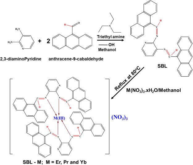
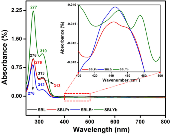
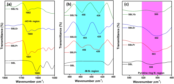
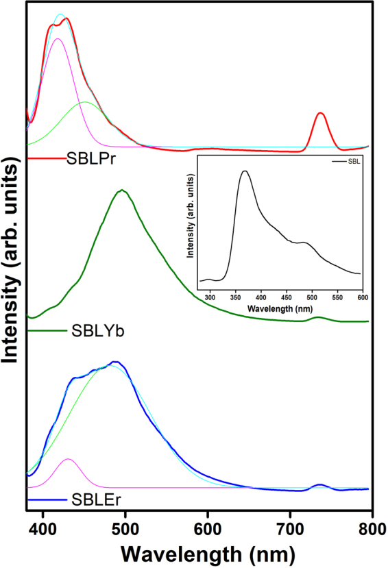
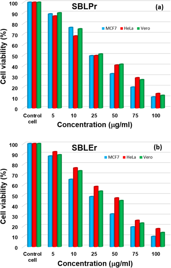
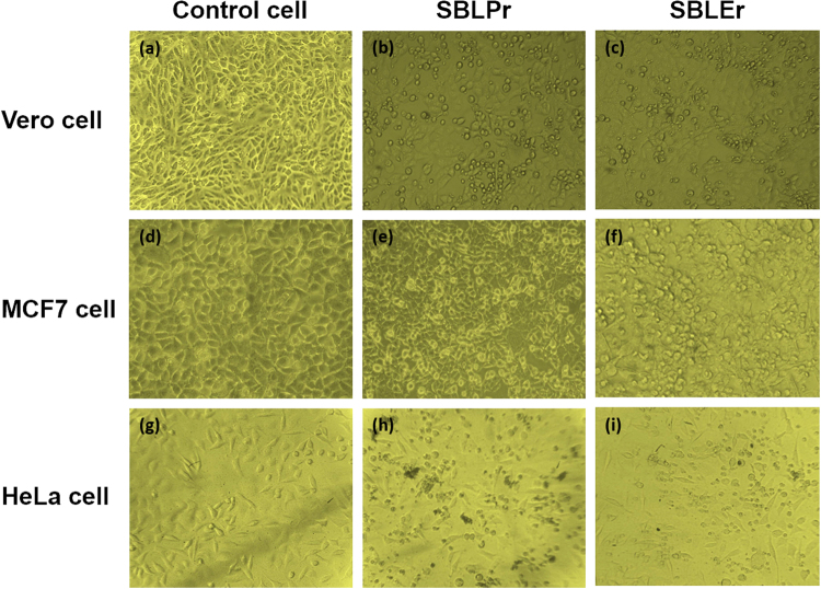
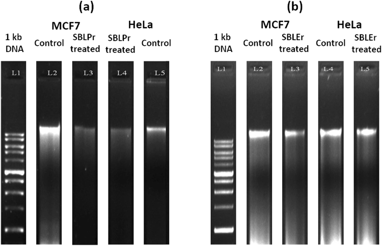
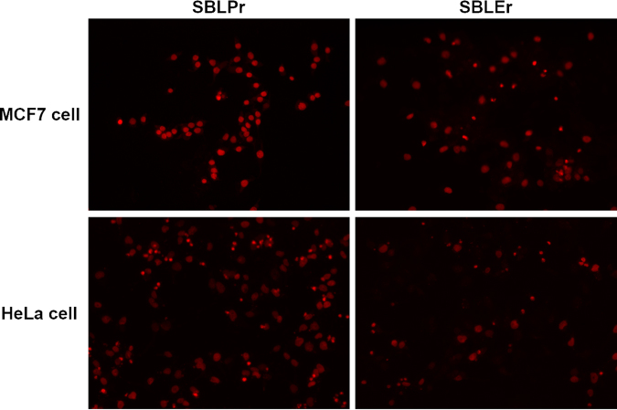
A Schiff base ligand (SBL), N2, N3-bis (anthracen-9-ylmethylene) pyridine-2, 3-diamine, was synthesized through the condensation of 2,6-diaminopyridine and anthracene-9-carbaldehyde using a 1:2 ratio. 1H NMR spectra confirmed the observation of non-involvement aromatic carboxylic proton in SBL. A novel series of lanthanide (i.e., praseodymium (Pr), erbium (Er), and ytterbium (Yb))-based SBL metal complexes was successfully synthesized, and their functional groups were elaborately demonstrated using UV-visible, Fourier transform infrared (FT-IR), and fluorescence spectroscopy analyses. FT-IR spectral studies revealed that SBL behaved as a bidentate ligand and it was structured with metal ions by the two azomethine nitrogens. The synthesized SBL-based metal complexes were elaborately performed for cytotoxicity activity versus Vero, human breast cancer (MCF7), and cervical (HeLa) anticancer cell lines.


Conflict of interest statement

The authors declare no competing interests.

Figures

Figure 1
Figure 1
Schematic methodology of SBL and its metal complex preparation.
Figure 2
Figure 2
UV-vis spectra of SBL and its metal complexes in range of 250–800 nm.
Figure 3
Figure 3
FT-IR spectra of SBL and its metal complexes. (a) Azomethine group (ν-HC=N) region, (b) -M-N-region, and (c) Pyridine ring N-region.
Figure 4
Figure 4
Fluorescence spectra of SBL metal complexes (Inset: fluorescence spectrum of SBL). The deconvoluted spectra included for SBLEr and SBLPr metal complexes were due to their combined broad spectral peak.
Figure 5
Figure 5
(a) TG and (b) DTA curves of SBL and its metal complexes.
Figure 6
Figure 6
SEM micrographs of (a) SBL and (b) SBLPr, (c) SBLEr, and (d) SBLYb complexes.
Figure 7
Figure 7
Cell viability of cancer cell lines (i.e., MCF7, HeLa, and Vero) against (a) SBLPr and (b) SBLEr metal complexes.
Figure 8
Figure 8
Morphological changes induced by SBLPr and SBLEr metal complexes using 25 μg/ml compared with control cancer cell lines.
Figure 9
Figure 9
(a) DNA fragmentation of MCF7 and HeLa IC50 cells treated with SBLPr complex at 24 h. Lane 1: 1 kb DNA ladder, Lane 2: MCF7 control DNA, Lane 3: SBLPr-treated MCF7 cell (25 μg/ml), Lane 4: SBLPr-treated HeLa cell (25 μg/ml), Lane 5: HeLa control DNA. (b) DNA fragmentation of MCF7 and HeLa IC50 cells treated with SBLEr complex at 24 h. Lane 1: 1 kb DNA ladder, Lane 2: MCF7 control DNA, Lane 3: SBLEr-treated MCF7 cell (25 μg/ml), Lane 4: HeLa control DNA, Lane 5: SBLEr-treated HeLa cell (25 μg/ml).
Figure 10
Figure 10
Morphological variations of AO/EB double-stained MCF7 and HeLa cells and their SBLPr- and SBLEr-treated cells using 25 μg/ml for 24 h.
Figure 11
Figure 11
Morphological variations of PI-stained MCF7 and HeLa cells after treated with SBLPr and SBLEr using 25 μg/ml for 24 h.

References

    1. Johnstone TC, Suntharalingam K, Lippard SJ. The Next Generation of Platinum Drugs: Targeted Pt(II) Agents, Nanoparticle Delivery, and Pt(IV) Prodrugs. Chem. Rev. 2016;116:3436–3486. doi: 10.1021/acs.chemrev.5b00597. - DOI - PMC - PubMed
    1. Woollins JD, Woollins A, Rosenberg B. The detection of trace amounts of trans-Pt(NH3)2Cl2 in the presence of cis-Pt(NH3)2Cl2. A high performance liquid chromatographic application of kurnakow’s test. Polyhedron. 1983;2:175–178. doi: 10.1016/S0277-5387(00)83954-6. - DOI
    1. Levi JA, Aroney RS, Dalley DN. Haemolytic anaemia after cisplatin treatment. Br. Med. J. 1981;282:2003–2004. doi: 10.1136/bmj.282.6281.2003. - DOI - PMC - PubMed
    1. Fuertes MA, Alonso C, Pérez JM. Biochemical Modulation of Cisplatin Mechanisms of Action: Enhancement of Antitumor Activity and Circumvention of Drug Resistance. Chem. Rev. 2003;103:645–662. doi: 10.1021/cr020010d. - DOI - PubMed
    1. Kathiresan S, Mugesh S, Annaraj J, Murugan M. Mixed-ligand copper(II) Schiff base complexes: the vital role of co-ligands in DNA/protein interactions and cytotoxicity. New J. Chem. 2017;41:1267–1283. doi: 10.1039/C6NJ03501A. - DOI

Publication types