. 2018 Jan 31:9:76.
doi: 10.3389/fimmu.2018.00076. eCollection 2018.

The Immunome in Two Inherited Forms of Pulmonary Fibrosis

Affiliations

The Immunome in Two Inherited Forms of Pulmonary Fibrosis

Souheil El-Chemaly et al. Front Immunol. .

Abstract

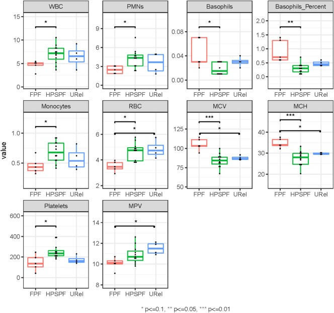
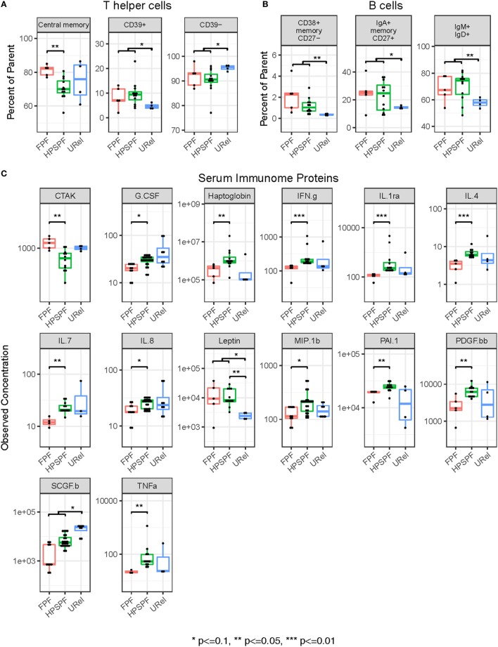
The immunome (immune cell phenotype, gene expression, and serum cytokines profiling) in pulmonary fibrosis is incompletely defined. Studies focusing on inherited forms of pulmonary fibrosis provide insights into mechanisms of fibrotic lung disease in general. To define the cellular and molecular immunologic phenotype in peripheral blood, high-dimensional flow cytometry and large-scale gene expression of peripheral blood mononuclear cells and serum proteomic multiplex analyses were performed and compared in a cohort with familial pulmonary fibrosis (FPF), an autosomal dominant disorder with incomplete penetrance; Hermansky-Pudlak syndrome pulmonary fibrosis (HPSPF), a rare autosomal recessive disorder; and their unaffected relatives. Our results showed high peripheral blood concentrations of activated central memory helper cells in patients with FPF. Proportions of CD38+ memory CD27- B-cells, IgA+ memory CD27+ B-cells, IgM+ and IgD+ B-cells, and CD39+ T helper cells were increased whereas those of CD39- T helper cells were reduced in patients affected with either familial or HPSPF. Gene expression and serum proteomic analyses revealed enrichment of upregulated genes associated with mitosis and cell cycle control in circulating mononuclear cells as well as altered levels of several analytes, including leptin, cytokines, and growth factors. In conclusion, dysregulation of the extra-pulmonary immunome is a phenotypic feature of FPF or HPSPF. Further studies investigating the blood immunome are indicated to determine the role of immune system dysregulation in the pathogenesis of pulmonary fibrosis.

Clinical trial registration: www.ClinicalTrials.gov, identifiers NCT00968084, NCT01200823, NCT00001456, and NCT00084305.

Keywords: B-cell; Hermansky–Pudlak syndrome; T-cell; cytokine; immunome; lymphocyte; pulmonary fibrosis; telomere disease.


Figures

Figure 1
Figure 1
Clinical images in patients with familial pulmonary fibrosis (FPF), Hermansky–Pudlak syndrome pulmonary fibrosis (HPSPF), and unaffected relatives (URels). High-resolution computed tomography scan images demonstrate lung fibrosis (white arrows) in patients with FPF (A) or HPSPF (B), and not in an URel (C). Whole mount electron microscopy images show absent platelet delta granules in a patient with Hermansky–Pudlak syndrome (D), size bar = 1 μm; normal control platelets contain delta granules (black arrows) (E), size bar = 0.5 μm.
Figure 2
Figure 2
Principal component analysis (PCA) of peripheral blood cells and serum immunome proteins in patients with familial pulmonary fibrosis (FPF), Hermansky–Pudlak syndrome pulmonary fibrosis (HPSPF), and unaffected relatives (URels). PCA of complete blood cell parameters shows clustering of patients with FPF, HPSPF, and URel (A). Subpopulations of patient groups are not found on PCA of peripheral blood mononuclear cell flow cytometry data (B). Patient groups also cluster with PCA of serum immunome proteomic data; one patient with FPF and their unaffected sibling segregate together as outliers (C).
Figure 3
Figure 3
Phenotyping of peripheral blood cell parameters in patients with familial pulmonary fibrosis (FPF), Hermansky–Pudlak syndrome pulmonary fibrosis (HPSPF), and unaffected relatives (URels). Concentrations of white blood cell (WBC), polymorphonuclear leukocyte (PMNs), basophils, monocytes, and platelets as well as basophil percent were significantly different in patients with FPF compared with those with HPSPF. Red blood cell (RBC) counts, mean corpuscular volume (MCV), mean corpuscular hemoglobin (MCH), and mean platelet volume (MPV) were also significantly different between groups as shown.
Figure 4
Figure 4
Flow cytometry of peripheral blood mononuclear cell populations and serum immunome profile in patients with familial pulmonary fibrosis (FPF), Hermansky–Pudlak syndrome pulmonary fibrosis (HPSPF), and unaffected relatives (URel). Percentages of central memory helper cells were significantly higher in patients with FPF compared with patients with HPSPF (A). Percentages of CD38+ memory CD27 B-cells, IgA+ memory CD27+ B-cells, IgM+ and IgD+ B-cells, CD39+ T helper cells, and CD39 T helper cells were significantly different in patients affected with either FPF or HPSPF compared with URel (A,B). Serum concentrations of chemokine ligand-27 (CTAK), granulocyte colony-stimulation factor (G.CSF), haptoglobin, interferon-γ (IFN.g), interleukin-1rα (IL.1ra), interleukin-4 (IL.4), interleukin-7 (IL.7), interleukin-8 (IL.8), leptin, macrophage inflammatory protein 1β (MIP.1b), plasminogen activator inhibitor-1 (PAI.1), platelet-derived growth factor-bb (PDGF.bb), stem cell growth factor-β (SCGF.b), and tumor necrosis factor-α (TNFa) were significantly different between groups as shown (C).
Figure 5
Figure 5
Analysis of peripheral blood mononuclear cell microarray expression data in patients with familial pulmonary fibrosis (FPF), Hermansky–Pudlak syndrome pulmonary fibrosis (HPSPF), and unaffected relatives (URel). Heatmaps of relative expression intensities of 31 differentially expressed genes with false discovery rate-adjusted p-value <0.1 and absolute log2-fold change > 0.3 in any pairwise comparison between three groups are displayed (A). Significantly enriched blood transcription modules with adjusted p-value <10−3 in at least one of three pairwise comparisons are shown (B).

References

    1. Martinez FJ, Safrin S, Weycker D, Starko KM, Bradford WZ, King TE, Jr, et al. The clinical course of patients with idiopathic pulmonary fibrosis. Ann Intern Med (2005) 142:963–7.10.7326/0003-4819-142-12_Part_1-200506210-00005 - DOI - PubMed
    1. Gross TJ, Hunninghake GW. Idiopathic pulmonary fibrosis. N Engl J Med (2001) 345:517–25.10.1056/NEJMra003200 - DOI - PubMed
    1. Raghu G, Rochwerg B, Zhang Y, Garcia CA, Azuma A, Behr J, et al. An official ATS/ERS/JRS/ALAT clinical practice guideline: treatment of idiopathic pulmonary fibrosis. An update of the 2011 clinical practice guideline. Am J Respir Crit Care Med (2015) 192:e3–19.10.1164/rccm.201506-1063ST - DOI - PubMed
    1. El-Chemaly S, Ziegler SG, Calado RT, Wilson KA, Wu HP, Haughey M, et al. Natural history of pulmonary fibrosis in two subjects with the same telomerase mutation. Chest (2011) 139:1203–9.10.1378/chest.10-2048 - DOI - PMC - PubMed
    1. Newton CA, Batra K, Torrealba J, Kozlitina J, Glazer CS, Aravena C, et al. Telomere-related lung fibrosis is diagnostically heterogeneous but uniformly progressive. Eur Respir J (2016) 48:1710–20.10.1183/13993003.00308-2016 - DOI - PMC - PubMed

Publication types

MeSH terms

Associated data