

Case Reports
doi: 10.1002/ccr3.1365.
eCollection 2018 Feb.
Azygos continuation of the caudal vena cava with segmental aneurysm, lung lobe torsion and pulmonary thromboembolism in a dog
Affiliations
- PMID: 29445478
- PMCID: PMC5799650
- DOI: 10.1002/ccr3.1365
Item in Clipboard
Case Reports
Azygos continuation of the caudal vena cava with segmental aneurysm, lung lobe torsion and pulmonary thromboembolism in a dog
Clin Case Rep.
.
Abstract
This case highlights the management and diagnostic evaluation of a dog with two individually rare conditions (lung lobe torsion and vena cava aneurysm) that ultimately resulted in fatal pulmonary thromboembolism.
Keywords: Blood clot; congenital; embolism; vascular anomaly.
Figures
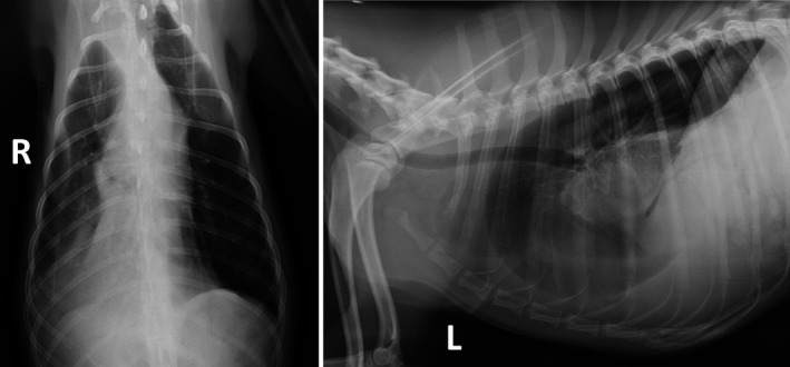
Ventrodorsal and left lateral thoracic radiographs, illustrating lung lobe torsion. Note the abnormal position of the right middle lung lobe along with a vesicular gas pattern and inability to visualize the right middle lobar bronchus. These findings are typical for lung lobe torsion.
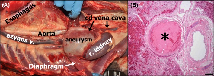
(A) Gross photograph of the thoracoabdominal cavity with most viscera removed, illustrating continuation of the abdominal caudal vena cava (with aneurysm) through the diaphragm as the azygos vein. The thoracic portion of the caudal vena cava, which terminated at the level of the liver, is not pictured as it was removed with the heart. (B) Photomicrograph of the lung depicting an occlusive pulmonary arterial thrombus (asterisk). A bronchus is located to the right of the thrombosed vessel, and hemorrhage is apparent in adjacent parenchyma to the left. Hematoxylin and eosin. Bar = 500 microns.
Similar articles
-
Resolution of Exercise-Induced Syncope After Stenting of the Azygos Vein in a Dog with Segmental Aplasia and Azygos Continuation of the Levopositioned Caudal Vena Cava.Animals (Basel). 2025 Mar 3;15(5):722. doi: 10.3390/ani15050722. Animals (Basel). 2025. PMID: 40076006 Free PMC article.
-
Computed tomographic and magnetic resonance imaging features of canine segmental caudal vena cava aplasia.J Small Anim Pract. 2009 Jul;50(7):341-9. doi: 10.1111/j.1748-5827.2009.00748.x. J Small Anim Pract. 2009. PMID: 19575698
-
Congenital absence of inferior vena cava as a rare cause of pulmonary thromboembolism.Yonsei Med J. 2004 Oct 31;45(5):947-51. doi: 10.3349/ymj.2004.45.5.947. Yonsei Med J. 2004. PMID: 15515211
-
[Duplication of the vena cava inferior with a continuation into the vena azygos. A report of a rare case].Minerva Chir. 1999 Apr;54(4):261-5. Minerva Chir. 1999. PMID: 10380526 Review. Italian.
-
[Congenital anomaly of the inferior vena cava with hemiazygos continuation. Ultrasonic diagnosis].Ann Radiol (Paris). 1990;33(6):339-46. Ann Radiol (Paris). 1990. PMID: 2085271 Review. French.
Cited by
-
Caudal vena cava aneurysm in a cat with Eisenmenger Syndrome.J Vet Med Sci. 2020 Jun 24;82(6):784-787. doi: 10.1292/jvms.19-0518. Epub 2020 May 15. J Vet Med Sci. 2020. PMID: 32418943 Free PMC article.
-
Single Posthepatic Portosystemic Shunt Communicated With Internal Thoracic Vein and Azygos Continuation of the Caudal Vena Cava in a Dog.Vet Med Sci. 2024 Nov;10(6):e70057. doi: 10.1002/vms3.70057. Vet Med Sci. 2024. PMID: 39315722 Free PMC article.
-
Dynamic Obstruction of an Anomalous Cavoazygos Vessel Associated with Interrupted Caudal Vena Cava in a Boxer Dog.CASE (Phila). 2021 Dec 16;6(1):36-42. doi: 10.1016/j.case.2021.11.005. eCollection 2022 Feb. CASE (Phila). 2021. PMID: 35243199 Free PMC article.
-
Situs inversus totalis with single extrahepatic portosystemic shunt and azygos continuation of the caudal vena cava in a dog: a case report.BMC Vet Res. 2025 Feb 22;21(1):87. doi: 10.1186/s12917-025-04565-7. BMC Vet Res. 2025. PMID: 39987442 Free PMC article.
-
CT findings of an incidental segmental aplasia of the caudal vena cava with azygos continuation in a guinea pig (Cavia porcellus).Open Vet J. 2024 Apr;14(4):1076-1080. doi: 10.5455/OVJ.2024.v14.i4.15. Epub 2024 Apr 30. Open Vet J. 2024. PMID: 38808297 Free PMC article.
References
-
- Alicioglu, B. , Kaplan M., and Ege T.. 2009. Absence of infrarenal inferior vena cava is not a congenital abnormality. Bratisl. Lek. Listy 110:304–306. - PubMed
-
- May, N. D. S. 1960. Absence of the prerenal segment of the posterior vena cava of the dog. Aust. Vet. J. 36:67–68.
-
- Laborda, J. , Gimeno M., Dominguez L., and Gill J.. 1996. Anomalous caudal vena cava in the dog. Vet. Rec. 138:20–21. - PubMed
-
- Barthez, P. Y. , Siemens L. M., and Koblik P. D.. 1996. Azygos continuation of the caudal vena cava in a dog: radiographic and ultrasonographic diagnosis. Vet. Radiol. Ultrasound. 37:354–356.
-
- Schwarz, T. , Rossi F., Wray J. D., Ablad B., Beal M. W., Kinns J., et al. 2009. Computed tomographic and magnetic resonance imaging features of canine segmental caudal vena cava aplasia. J. Small Anim. Pract. 50:341–349. - PubMed
Publication types
LinkOut - more resources
Full Text Sources
Other Literature Sources
