. 2018 Feb 6;5(1):ENEURO.0071-17.2018.
doi: 10.1523/ENEURO.0071-17.2018. eCollection 2018 Jan-Feb.

In Vivo Observation of Structural Changes in Neocortical Catecholaminergic Projections in Response to Drugs of Abuse

Affiliations

In Vivo Observation of Structural Changes in Neocortical Catecholaminergic Projections in Response to Drugs of Abuse

Mai M Morimoto et al. eNeuro. .

Abstract

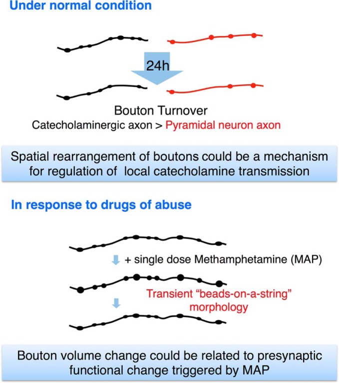
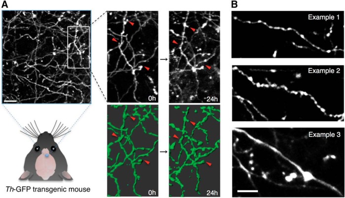
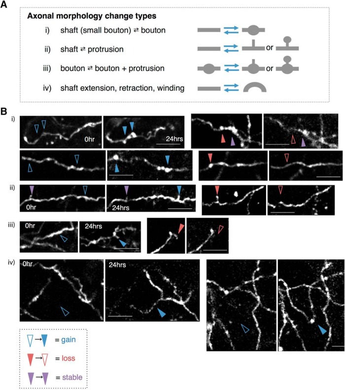
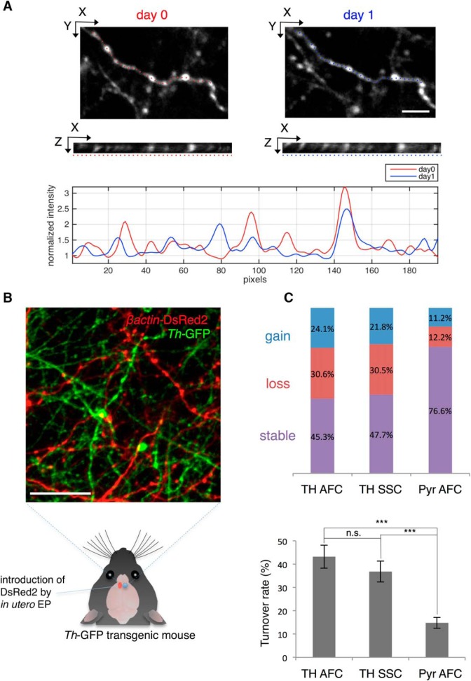
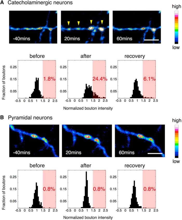
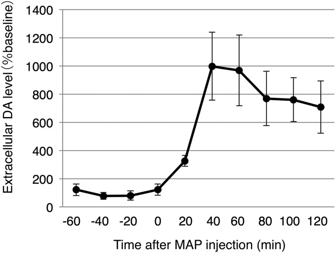
Catecholaminergic (dopamine and norepinephrine) projections to the cortex play an important role in cognitive functions and dysfunctions including learning, addiction, and mental disorders. While dynamics of glutamatergic synapses have been well studied in such contexts, little is known regarding catecholaminergic projections, owing to lack of robust methods. Here we report a system to monitor catecholaminergic projections in vivo over the timeframes that such events occur. Green fluorescent protein (GFP) expression driven by tyrosine hydroxylase promoter in a transgenic mouse line enabled us to perform two-photon imaging of cortical catecholaminergic projections through a cranial window. Repetitive imaging of the same axons over 24 h revealed the highly dynamic nature of catecholaminergic boutons. Surprisingly, administration of single high dose methamphetamine (MAP) induced a transient increase in bouton volumes. This new method opens avenues for longitudinal in vivo evaluation of structural changes at single release sites of catecholamines in association with physiology and pathology of cortical functions.

Keywords: axon; catecholamine; in vivo imaging; methamphetamine; neocortex; transgenic mouse.


Figures

None
Graphical abstract
Figure 1.
Figure 1.
In vivo imaging of catecholaminergic axons labeled by Th-GFP transgenic line in the mouse neocortex. A, In vivo two-photon image of catecholaminergic axons observed in Th-GFP transgenic mice. The same axons were repeatedly observed in following experiments. Grayscale images show Z-projections of image stack. Axons in green are 3D-rendered versions. Red arrowheads indicate change events. Scale bar: 15 μm. B, Representative morphology of the major three examples of catecholaminergic axons observed in Th-GFP transgenic mice. Most axons were thin and varicose, except for occasional large smooth fibers in deeper layers (∼100 μm). Scale bar: 10 μm.
Figure 2.
Figure 2.
Morphologic changes of catecholaminergic axons over 24 h. A, Types of morphologic changes observed between the two time points (0 and 24 h). B, Examples of the grouped changes i–iv. Blue, red, and purple arrowheads indicate examples of gained, lost, and stable components, respectively.
Figure 3.
Figure 3.
Bouton dynamics of catecholaminergic axons and pyramidal neurons over 24 h. A, Quantification of bouton dynamics over 24 h. Well-isolated axonal arbors were selected for analysis (top: XY plane, middle: XZ plane along the axon; dotted line). Gained, lost, and stable boutons were scored based on the pixel intensity profile along the axon (bottom). Scale bar: 5 μm. B, Observation of Layer II/III pyramidal neuron axons (red) and catecholaminergic axons (green) within the same preparation in vivo. Scale bar: 25 μm. C, Top, Fraction of gained, lost, and stable boutons of dopaminergic axons in the AFC (TH AFC), catecholaminergic axons in the SSC (TH SSC), and pyramidal neuron axons in the AFC (Pyr AFC). TH AFC: N = 5, n = 232; TH SSC: N = 5, n = 197; Pyr AFC: N = 5, n = 278. Bottom, Comparison of TORs between axon types. N: number of animals, n: number of boutons. Data are presented as mean ± SEM; ***p < 0.001, n.s.: not significant.
Figure 4.
Figure 4.
Morphologic change of catecholaminergic boutons after MAP injection in the AFC. A single dose of MAP (10 mg/kg) induced a transient beads-on-a-string morphology in catecholaminergic neurons (A), while in pyramidal neurons, there was no obvious structural change (B); yellow arrowheads: enlarged boutons; scale bar: 10 μm. The histograms show bouton intensity normalized to MAP injection time point. After injection, the distribution of catecholaminergic bouton intensity shifted to above 1.5 times its original intensity (light red background), while pyramidal neuron boutons did not. Catecholamine neurons: N = 5, n = 393; pyramidal neurons: N = 5, n = 263 (N: number of animals, n: number of boutons).
Figure 5.
Figure 5.
Extracellular dopamine concentration measured by microdialysis in the AFC. Mice were kept under anesthesia in an identical condition to the in vivo imaging experiments. A single dose of MAP (10 mg/kg) was injected at time 0 and was monitored in 20 min intervals. N = 4 mice.

Similar articles

Cited by

References

    1. Akil M, Pierri JN, Whitehead RE, Edgar CL, Mohila C, Sampson AR, Lewis DA (1999) Lamina-specific alterations in the dopamine innervation of the prefrontal cortex in schizophrenic subjects. Am J Psychiatry 156:1580–1589. 10.1176/ajp.156.10.1580 - DOI - PubMed
    1. Arnsten AF (1998) Catecholamine modulation of prefrontal cortical cognitive function. Trends in Cogn Sci 2:436–447. 10.1016/S1364-6613(98)01240-6 - DOI - PubMed
    1. Berger B, Thierry AM, Tassin JP, Moyne MA (1976) Dopaminergic innervation of the rat prefrontal cortex: a fluorescence histochemical study. Brain Res 106:133–145. 10.1016/0006-8993(76)90078-0 - DOI - PubMed
    1. Bubser M, Schmidt WJ (1990) 6-Hydroxydopamine lesion of the rat prefrontal cortex increases locomotor activity, impairs acquisition of delayed alternation tasks, but does not affect uninterrupted tasks in the radial maze. Behav Brain Res 37:157–168. 10.1016/0166-4328(90)90091-R - DOI - PubMed
    1. Cubells JF, Rayport S, Rajendran G, Sulzer D (1994) Methamphetamine neurotoxicity involves vacuolation of endocytic organelles and dopamine-dependent intracellular oxidative stress. J Neurosci 14:2260–2271. - PMC - PubMed

Publication types

LinkOut - more resources