Review
. 2019 Jan 10;62(1):3-23.
doi: 10.1021/acs.jmedchem.7b01640. Epub 2018 Mar 5.

Positive and Negative Allosteric Modulators of N-Methyl-d-aspartate (NMDA) Receptors: Structure-Activity Relationships and Mechanisms of Action

Affiliations
Review

Positive and Negative Allosteric Modulators of N-Methyl-d-aspartate (NMDA) Receptors: Structure-Activity Relationships and Mechanisms of Action

Erica S Burnell et al. J Med Chem. .

Abstract

Excitatory activity in the CNS is predominately mediated by l-glutamate through several families of l-glutamate neurotransmitter receptors. Of these, the N-methyl-d-aspartate receptor (NMDAR) family has many critical roles in CNS function and in various neuropathological and psychiatric conditions. Until recently, the types of compounds available to regulate NMDAR function have been quite limited in terms of mechanism of action, subtype selectivity, and biological effect. However, several new classes of NMDAR agents have now been identified that are positive or negative allosteric modulators (PAMs and NAMs, respectively) with various patterns of NMDAR subtype selectivity. These new agents act at several newly recognized binding sites on the NMDAR complex and offer significantly greater pharmacological control over NMDAR activity than previously available agents. The purpose of this review is to summarize the structure-activity relationships for these new NMDAR modulator drug classes and to describe the current understanding of their mechanisms of action.


Figures

Figure 1
Figure 1
NMDA receptor structure. (Top) Crystal structure of the tetrameric NMDA receptor complex of Xenopus laevis without intracellular C-terminals (PDB: 4TLM). The two GluN1 backbones are shown in green/blue and the two GluN2B backbones are in yellow/red. (Bottom row) A single GluN1 subunit in the same orientation as the blue GluN1 subunit in the top, left panel. Middle panel showing this subunit’s secondary structure (beta sheets, blue / alpha helix, red) and right panel: stick representation of backbone and side chains with the ATD - green, S1 and linker to M1- red, S2 and linker to M4 - blue, M1 – yellow, M2 - orange, M3 - magenta, and M4 - cyan. The three domain levels are shown on the left for ATD, LBD, and transmembrane (TM) domain.
Figure 2:
Figure 2:
(a) Lead compounds 1 (PS) and 2 (PAS); (b) General SAR; (c) NAM 3.
Figure 3:
Figure 3:
Structures of cholesterol (4), 24(S)-HC (5), SGE-201 (6) and 25-HC (7).
Figure 4:
Figure 4:
(a) Structure of lead compound 8 (TCN-201); (b) compound 9; (c) Most selective compound for GluN2A (9, MPX-004) and most potent compound at GluN2A (10, MPX-007).
Figure 5
Figure 5
Allosteric modulator binding sites on the NMDA receptor. (Left panel) Crystal structure of the tetrameric NMDA receptor complex of Xenopus laevis without intracellular C-terminals (PDB: 4TLM). The two GluN1 backbones are shown in green/blue and the two GluN2B backbones are in yellow/red. Homologous amino acid residues which are involved in the binding and/or actions of allosteric modulators are shown as space-filled: PYD-1 – light blue; GNE-6901/TCN-201 - yellow; UBP512 - pink; QNZ46/DQP-1105 - orange; GNE-9278 - green; CIQ - purple. Also shown are space-filled ligands with CPK coloring: Ro25–6981 bound in the ATD, glycine-site agonist 1-aminocyclopropanecarboxylic acid bound in the GluN1 LBD, and glutamate-site agonist trans-1-aminocyclobutane-1,3-dicarboxylic acid bound in the GluN2B LBD. (Right) LBD dimer with homologous residues that interact with TCN-201 are shown as space-filled in blue (GluN1) and yellow (GluN2). Bound glycine and L-glutamate are shown in CPK coloring.
Figure 6
Figure 6
(A,B) GluN1/GluN2A LBD dimer with bound TCN-201 (yellow), L-glutamate (green), and glycine (light blue) PDB: 5I56 . (C) TCN-201 binding pocket with interacting residues shown in stick mode.
Figure 7:
Figure 7:
(a) Lead compounds 12 and 13; (b) CP 465022 (14); (c) General SAR observations; (d) Representative compounds 15 (QNZ46) and 16.
Figure 8
Figure 8
Allosteric sites associated in or near the TM domain. The two GluN1 backbones are shown in green/blue and the two GluN2B backbones are in yellow/red. Homologous amino acid residues which are involved in the binding and/or actions of allosteric modulators are shown as space-filled: UBP512 - pink; QNZ46/DQP-1105 - orange; GNE-9278 - green; CIQ - purple. UBP512 and PS activity also involves residues in the S2 domain (not shown).
Figure 9:
Figure 9:
(a) Lead compound 17; (b) General SAR observations; (c) Representative compounds 18 (DQP-26) and 19 (DQP-1105).
Figure 10:
Figure 10:
(a) Lead compound 20; (b) General SAR observations; (c) Compounds 21 and 22.
Figure 11:
Figure 11:
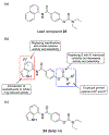
(a) Lead compound 23; (b) General SAR observations; (c) Compound 24 (NAB-14).
Figure 12:
Figure 12:
(a) Lead compound 25; (b) General SAR observations; (c) 26 (UBP710), 27 (UBP646).
Figure 13:
Figure 13:
(a) General SAR observations; (c) Representative naphthalene derivatives from the series: 28 (UBP684), 29 (UBP618), 30 (UBP551) and 31 (UBP552).
Figure 14:
Figure 14:
(a) Lead compound 32 (UBP608); (b) General SAR observations; (c) Representative coumarin derivatives from the series: 33 (UBP656) and 34 (UBP714).
Figure 15:
Figure 15:
Structure of GNE-9278 (35).
Figure 16:
Figure 16:
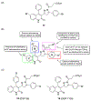
(a) Lead compound GNE 3476 (36); (b) Summary of SAR study; (c) Structures of GNE-0723 (37), GNE-5729 (38), GNE-6901 (39) and GNE-8324 (40).
Figure 17:
Figure 17:
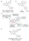
(a) Lead compound 41; (b) General structure for SAR studies; (c) Compounds 42 (PYD-111) and 43 (PYD-106).
Figure 18
Figure 18
Residues important for PYD-106 GluN1/GluN2C-selective PAM activity. (Left) Residues important for NAM/PAM activity are shown in the tetrameric complex with GluN1 backbones in blue/green and GluN2 backbones in red/yellow (PDB: 4TLM). Residues homologous to those important for PYD-106 GluN2C-selectivity are shown on a single GluN2 structure as space-filled in light blue. These sites oppose each other across the ATD/LBD interface.
Figure 19:
Figure 19:
(a) Lead compound 44 (CIQ); (b) General SAR observations; (c) Compound 45.
Figure 20:
Figure 20:
(a) Lead GluN2B/C/D PAM 46; (b) General SAR observations; (c) Active enantiomer of 46.

References

    1. Watkins JC; Evans RH Excitatory amino acid transmitters. Annu Rev Pharmacol Toxicol 1981, 21, 165–204. - PubMed
    1. Monaghan DT; Bridges RJ; Cotman CW The excitatory amino acid receptors: their classes, pharmacology, and distinct properties in the function of the central nervous system. Annu Rev Pharmacol Toxicol 1989, 29, 365–402. - PubMed
    1. Dingledine R; Borges K; Bowie D; Traynelis SF The glutamate receptor ion channels. Pharmacol Rev 1999, 51, 7–61. - PubMed
    1. Traynelis SF; Wollmuth LP; McBain CJ; Menniti FS; Vance KM; Ogden KK; Hansen KB; Yuan H; Myers SJ; Dingledine R Glutamate receptor ion channels: structure, regulation, and function. Pharmacol Rev 2010, 62, 405–496. - PMC - PubMed
    1. Paoletti P; Bellone C; Zhou Q NMDA receptor subunit diversity: impact on receptor properties, synaptic plasticity and disease. Nat Rev Neurosci 2013, 14, 383–400. - PubMed

MeSH terms

LinkOut - more resources