Comparative Study
. 2018 Feb 15;13(2):e0193006.
doi: 10.1371/journal.pone.0193006. eCollection 2018.

Differences between fellow eyes of acute and chronic primary angle closure (glaucoma): An ultrasound biomicroscopy quantitative study

Affiliations
Comparative Study

Differences between fellow eyes of acute and chronic primary angle closure (glaucoma): An ultrasound biomicroscopy quantitative study

Mengwei Li et al. PLoS One. .

Abstract

Purpose: To compare various biometric parameters between fellow eyes of acute primary angle closure (glaucoma) [APAC(G)] and fellow eyes of chronic primary angle closure (glaucoma) [CPAC(G)].

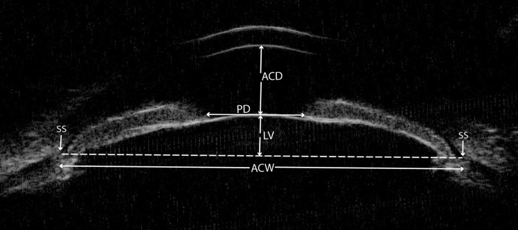
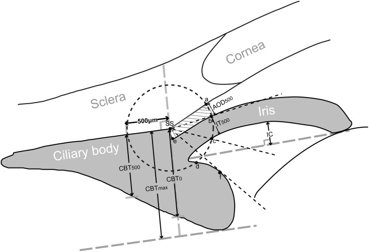
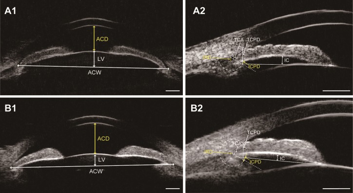
Methods: Ultrasound biomicroscopy examinations were performed on 47 patients with unilateral APAC(G) and 41 patients with asymmetric CPAC(G) before laser peripheral iridotomy and pilocarpine treatment. Anterior chamber depth and width (ACD and ACW), lens vault (LV), iris curvature (IC), iris root distance (IRD), trabecular-ciliary process distance (TCPD), iris-ciliary process distance (ICPD), trabecular-ciliary angle (TCA), and other biometric parameters were compared between fellow eyes of APAC(G) and fellow eyes of CAPC(G).

Results: Compared with fellow eyes of CPAC(G), fellow eyes of APAC(G) had smaller ACD (P < 0.001), ACW (P = 0.007), TCPD (P = 0.016), ICPD (P = 0.008), and TCA (P = 0.006), as well as larger LV (P = 0.002), IC (P = 0.012), and IRD (P = 0.003). On multivariate logistic regression analyses, a 0.1 mm decrease in ACD (odds ratio [OR]: 0.705, 95%CI: 0.564-0.880, P = 0.002), ICPD (OR: 0.557, 95%CI: 0.335-0.925, P = 0.024), and a 0.1 mm increase in IRD (OR: 2.707, 95%CI: 1.025-7.149, P = 0.045), was significantly associated with occurrence of acute angle closures.

Conclusions: Fellow eyes of APAC(G) had smaller anterior segment dimensions, higher LV, more posterior iris insertion, greater IC, and more anteriorly rotated ciliary body compared with fellow eyes of CPAC(G). ACD, ICPD, and IRD were the three most important parameters that distinguish eyes predisposed to APAC(G) or CPAC(G).


Conflict of interest statement

Competing Interests: The authors have declared that no competing interests exist.

Figures

Fig 1
Fig 1. The determination of the parameters on an ultrasound biomicroscopy image of the horizontal perpendicular full view scans at the nasal-temporal position centered over the pupil.
ACD = anterior chamber depth; ACW = anterior chamber width; LV = lens vault; PD = pupil diameter; SS = scleral spur.
Fig 2
Fig 2. The determination of the parameters on an ultrasound biomicroscopy diagram of the radial scans centered over the limbus.
A circle with a radius of 500 μm centered on the scleral spur (SS) is drawn. Angle-opening distance at 500 μm (AOD500) is the distance between the posterior corneal surface and the anterior iris surface on a line perpendicular to the trabecular meshwork 500 μm from the SS. Trabecular-iris space area at 500 μm (TISA500) is the area bounded anteriorly by AOD500 as determined, posteriorly by a line drawn from the SS perpendicular to the plane of the inner scleral wall to the iris, superiorly by the inner corneoscleral wall, and inferiorly by the iris surface. Trabecular-anterior iris surface angle (TAIA) is the angle between the posterior corneal surface and the anterior iris surface (angle of “a-SS-b”). Trabecular-posterior iris surface angle (TPIA) is the angle between the posterior corneal surface and the posterior iris surface (angle of “a-SS-c”). Iris thickness at 500 μm (IT500) is iris thickness at 500 μm from the SS. Iris curvature (IC) is the perpendicular distance from a line between the most central to the most peripheral points of the iris pigment epithelium to the posterior iris surface at the point of greatest convexity. Iris root distance (IRD): the distance from the SS to the insertion location of the iris into the ciliary body (line of “SS-e”). Trabecular-ciliary process distance (TCPD) is a line extending from the corneal endothelium 500 μm anterior to the SS toward the ciliary processes (line of “ad”). Iris-ciliary process distance (ICPD) is the posterior surface of the iris 500 μm anterior to the SS toward the ciliary processes (line of “cd”). Trabecular-ciliary angle (TCA) is the angle between the posterior corneal surface and the anterior surface of the ciliary body (angle of “a-SS-f”). Maximum ciliary body thickness (CBTmax) is the distance from the most inner point of the ciliary body to the inner wall of sclera or its extended line. Ciliary body thicknesses at the point of the SS (CBT0) and at a distance of 500 μm (CBT500) are also measured.
Fig 3
Fig 3. Ultrasound biomicroscopy images of two patients (patient A and B).
A, The fellow eye of a patient with acute primary angle closure (APAC). B, The fellow eye of a patient with chronic primary angle closure (CPAC). Note that the fellow eye of APAC has smaller anterior segment dimensions (anterior chamber depth [ACD] and anterior chamber width [ACW]), higher lens vault (LV) (A1 vs. B1), greater iris curvature (IC), more posterior iris insertion (longer iris root distance [IRD]), and more anteriorly positioned ciliary body (shorter trabecular-ciliary process distance [TCPD] and iris-ciliary process distance [ICPD], and smaller trabecular-ciliary angle [TCA]) (A2 vs. B2). Scale bar: 1mm.

References

    1. Foster PJ, Johnson GJ. Glaucoma in China: How big is the problem? Br J Ophthalmol. 2001;85:1277–1282. doi: 10.1136/bjo.85.11.1277 - DOI - PMC - PubMed
    1. Sun X, Dai Y, Chen Y, Yu DY, Cringle SJ, Chen J, et al. Primary angle closure glaucoma: What we know and what we don't know. Prog Retin Eye Res. 2017;57:26–45. doi: 10.1016/j.preteyeres.2016.12.003 - DOI - PubMed
    1. Wright C, Tawfik MA, Waisbourd M, Katz LJ. Primary angle-closure glaucoma: An update. Acta Ophthalmol. 2016;94:217–225. doi: 10.1111/aos.12784 - DOI - PubMed
    1. Nongpiur ME, Ku JY, Aung T. Angle closure glaucoma: A mechanistic review. Curr Opin Ophthalmol. 2011;22:96–101. doi: 10.1097/ICU.0b013e32834372b9 - DOI - PubMed
    1. Nongpiur ME, He M, Amerasinghe N, Friedman DS, Tay WT, Baskaran M, et al. Lens vault, thickness, and position in Chinese subjects with angle closure. Ophthalmology. 2011;118:474–479. doi: 10.1016/j.ophtha.2010.07.025 - DOI - PubMed

Publication types

LinkOut - more resources