. 2018 Feb 15;14(2):e1006853.
doi: 10.1371/journal.ppat.1006853. eCollection 2018 Feb.

Dynamic remodeling of lipids coincides with dengue virus replication in the midgut of Aedes aegypti mosquitoes

Affiliations

Dynamic remodeling of lipids coincides with dengue virus replication in the midgut of Aedes aegypti mosquitoes

Nunya Chotiwan et al. PLoS Pathog. .

Abstract

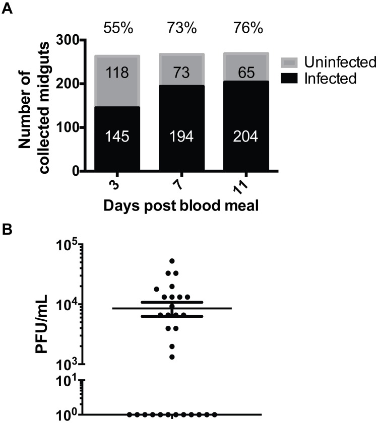
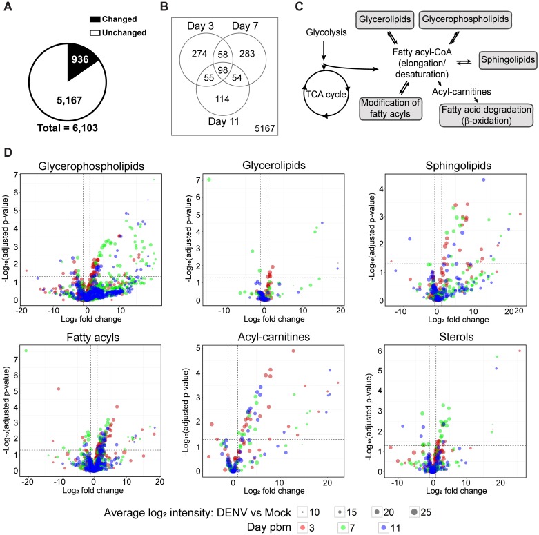
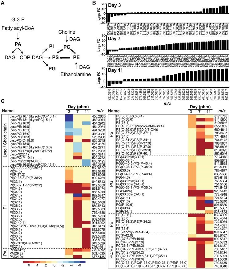
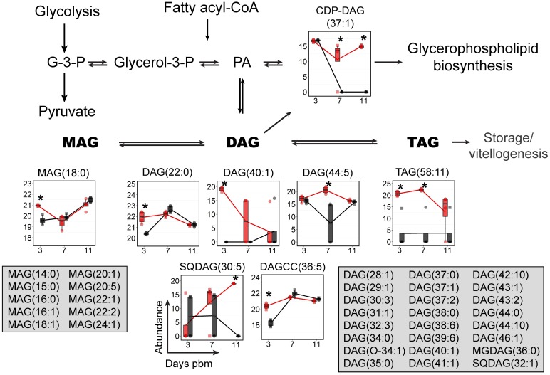
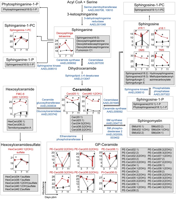
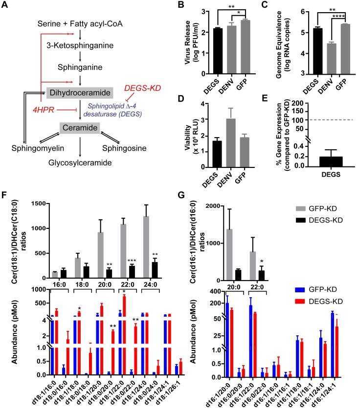
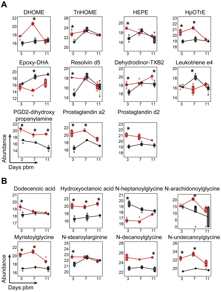
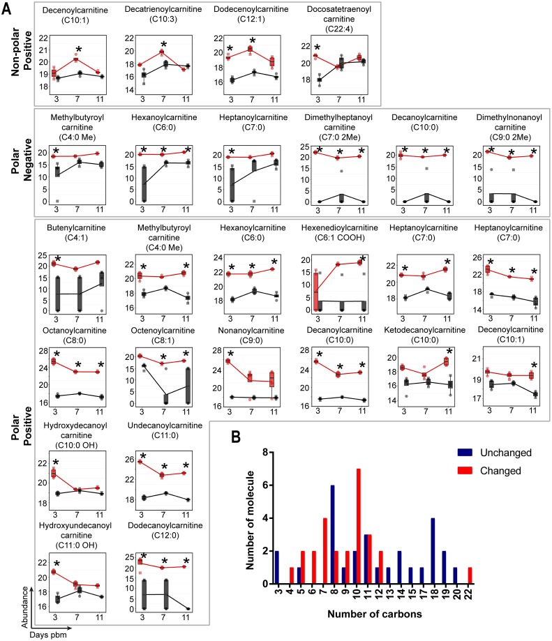
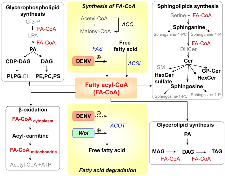
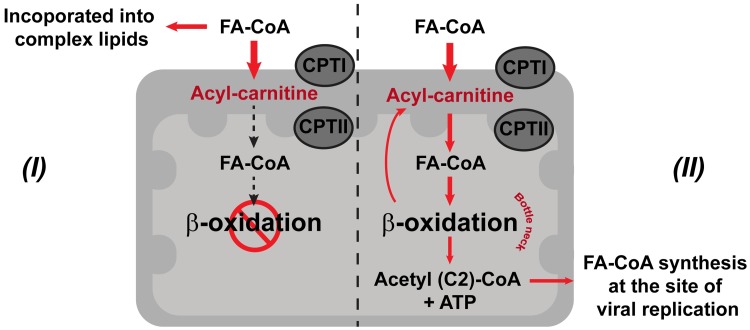
We describe the first comprehensive analysis of the midgut metabolome of Aedes aegypti, the primary mosquito vector for arboviruses such as dengue, Zika, chikungunya and yellow fever viruses. Transmission of these viruses depends on their ability to infect, replicate and disseminate from several tissues in the mosquito vector. The metabolic environments within these tissues play crucial roles in these processes. Since these viruses are enveloped, viral replication, assembly and release occur on cellular membranes primed through the manipulation of host metabolism. Interference with this virus infection-induced metabolic environment is detrimental to viral replication in human and mosquito cell culture models. Here we present the first insight into the metabolic environment induced during arbovirus replication in Aedes aegypti. Using high-resolution mass spectrometry, we have analyzed the temporal metabolic perturbations that occur following dengue virus infection of the midgut tissue. This is the primary site of infection and replication, preceding systemic viral dissemination and transmission. We identified metabolites that exhibited a dynamic-profile across early-, mid- and late-infection time points. We observed a marked increase in the lipid content. An increase in glycerophospholipids, sphingolipids and fatty acyls was coincident with the kinetics of viral replication. Elevation of glycerolipid levels suggested a diversion of resources during infection from energy storage to synthetic pathways. Elevated levels of acyl-carnitines were observed, signaling disruptions in mitochondrial function and possible diversion of energy production. A central hub in the sphingolipid pathway that influenced dihydroceramide to ceramide ratios was identified as critical for the virus life cycle. This study also resulted in the first reconstruction of the sphingolipid pathway in Aedes aegypti. Given conservation in the replication mechanisms of several flaviviruses transmitted by this vector, our results highlight biochemical choke points that could be targeted to disrupt transmission of multiple pathogens by these mosquitoes.


Conflict of interest statement

"The authors have declared that no competing interests exist."

Figures

Fig 1
Fig 1. DENV infection in mosquitoes.
Individual midguts dissected from DENV-fed mosquitoes (n = 799) were tested for the presence of viral RNA by qRT-PCR. (A) Percent of virus-RNA positive midguts are shown above the bar graphs. (B) Infectious viral titers of a representative subset (n = 30) of whole mosquitoes harvested on day 11 pbm were determined to evaluate the range of titers observed in the experiment following dissemination.
Fig 2
Fig 2. Metabolic profile of the mosquito midgut during the course of DENV infection.
Significant changes were observed in the metabolic profile of the midgut upon infection. (A) The pie chart shows numbers of features in the midgut detected on days 3, 7 and 11 following a DENV-infectious blood meal compared to a noninfectious blood meal with significantly altered levels of abundance (|log2 fold change| ≥ 1 and p-value < 0.05) in black and nonsignificantly altered levels of abundance (|log2 fold change| < 1 or p-value ≥ 0.05) in white. (B) Venn diagram shows numbers of features that were altered in abundance in DENV-infected midguts compared to uninfected midguts on the days 3, 7, and 11 pbm. (C) Overview of lipid classes observed in this study and their relationship to each other within metabolic pathways. Fatty acyl-CoAs can be de novo synthesized from intermediates in central carbon metabolism. They can be further modified or incorporated into several classes of more complex lipids or converted to acyl-carnitine for energy production. (D) Volcano plots show the abundances of metabolites from different classes of lipids detected on days 3, 7 and 11 pbm in DENV-infected versus uninfected midguts. The vertical dashed lines indicate a 2-fold change in abundance and the horizontal dashed line indicates a p-value = 0.05.
Fig 3
Fig 3. GP fluctuation following DENV infection of mosquito midguts.
(A) The metabolic pathway linking GP classes. Highlighted in bold are the GPs observed in this study. (B) GP landscape altered upon DENV infection in midguts. GP species that were altered on days 3, 7, and 11 pbm are listed by m/z and arranged by Log2 fold changes from lowest to highest. A significant burst of GP abundance was observed at day 7 post-infection coinciding with increased viral replication in the midgut. (C) Heatmap of Log2 fold changes of GP species on days 3, 7 and 11 arranged by putative ID subclasses. Abbreviations: CDP-DAG, cytidine diphosphate diacylglycerol; DAG, diacylglycerol; G-3-P, glyceraldehyde-3-phosphate; PA, phosphatidic acid; PC, phosphatidylcholine; PE, phosphatidylethanolamine; PG, phosphatidylglycerol; PI, phosphatidylinositol; PS, phosphatidylserine.
Fig 4
Fig 4. GL levels were dynamically altered upon DENV infection of mosquito midguts.
The abundances of GL species detected in mosquito midguts were mapped to the GL biosynthesis pathway. GL molecules that were significantly altered in abundance are shown in boxplots. Feature that were detected but did not change in abundance are listed in the grey boxes. The abundance of metabolites detected in DENV-infected samples is shown in red and uninfected samples in black. Individual sample pools are represented by circles and squares and technical replicates are dots with the same symbol. Asterisk (*) indicates significantly different levels of abundance between DENV-infected and uninfected samples (|log2 fold change| ≥ 1, p < 0.05) Abbreviations: DAG, diacylglycerol; CDP-DAG, cytidine diphosphate diacylglycerol; G-3-P, glyceraldehyde-3-phosphate; glycerol-3-P, glycerol-3-phosphate; MAG, monoacylglycerol; PA, phosphatidic acid; TAG, triacylglycerol; SQDAG, Sulfoquinovosyldiacylglycerol and DAGCC, diacylglycerylcarboxy-N-hydroxymethyl-choline.
Fig 5
Fig 5. Accumulation of SPs during DENV infection.
The SP pathway for Ae. aegypti was reconstructed for the first time in this study. This SP metabolic pathway (modified from Hannun et al., 2008 [38]) is shown overlaid with box plots with SPs that exhibited significantly altered levels during infection. Features that were detected but unchanged in abundance upon DENV infection are listed in the grey boxes. The abundance of metabolites detected in DENV-infected samples is shown in red and uninfected samples in black. Individual sample pools are represented by circles and squares and technical replicates are dots with the same symbol. Asterisks (*) indicates significantly different levels of abundance between DENV-infected and uninfected samples (|log2 fold change| ≥ 1, p < 0.05). The enzymes predicted to catalyze the reactions in the SP pathway and their VectorBase accession numbers (AAELXXXXXX) according to AaegEL5 assembly and as annotated in the KEGG pathway are given in blue text. Where enzymes are not given it indicates an absence of annotation for Ae. aegypti of names via the KEGG database and VectorBase. Abbreviations: Cer, ceramide; GP-Cer, glycerophospholipid-ceramide; HexCer, hexosylceramide; FMC-6, acetyl-sphingosine-tetraacetyl-GalCer(d40:1(2OH)); PE-Cer, phosphatidylethanolamine-ceramide; Phytosphinganine-1-P, phytosphinganine-1-phosphate; PI-Cer, phosphatidylinositol-ceramide; Sphingosine-1-P, sphingosine-1-phosphate; Sphingosince-1-PC, sphingosine-1-phosphatidylcholine and SM, sphingomyelin.
Fig 6
Fig 6. Changes in the Cer-DHCer balance impair DENV infection in Aag2 cells.
(A) A schematic of the SP biosynthesis pathway showing the precursor (DHCer) and product (Cer) of the sphingolipid Δ-4 desaturase (DEGS) enzyme-catalyzed reaction. In this experiment, the Cer/DHCer balance was changed by dsRNA knockdown of the DEGS gene expression as well as pharmacological inhibition of DEGS with 4HPR (S2 Fig). (B and C) Aag2 cells were transfected with dsRNA derived from DEGS, DENV (positive control) or GFP (negative control) genes. Two days post transfection, cells were infected with DENV at 0.3 MOI. Cell culture supernatant (B) or intracellular total RNA (C) were collected at 24 hours post infection (hpi) and analyzed for infectious virus in medium and intracellular DENV RNA, respectively. One-way ANOVA followed by Dunnett’s multiple comparisons test were used for statistical analysis. (D) Viability of Aag2 cells after dsRNA treatments at 48 h post dsRNA transfection. (E) DEGS mRNA expression in DEGS-KD cells compared to the expression of the DEGS mRNA in GFP-KD cells (set at a 100%). (F and G) Analysis of Cer and DHCer species in DEGS-KD (red) and GFP-KD cells (blue) using LC-MS/MS Multiple Reaction Monitoring (N = 3). (F) and (G) shows Cer and DHCer molecules with 18- and 16-carbon long chain sphingoid bases, respectively. Lower panels show the abundance of individual species of Cer(d18:1/xx:x or d16:1/xx:x) or DHCer(d18:0/xx:x or d16:0/xx:x) where xx:x refers to the fatty acyl chain attached to each sphingoid head group. The upper panels show the ratio of Cer/DHCer species with the same fatty acyl chain lengths. These ratios highlight the Cer-DHCer imbalance caused by manipulation of DEGS gene expression. Student’s t-test was applied for statistical analysis. * p < 0.05, ** p < 0.01, *** p < 0.005, **** p < 0.001.
Fig 7
Fig 7. Temporal fluctuations in levels of fatty acyl molecules following DENV infection of mosquito midguts.
(A) The abundances of fatty acyls that have known functions in immunomodulation. (B) The abundances of free fatty acids and N-acylamides that have known functions in signaling and/or that are markers of malfunction in fatty acid oxidation are shown. The abundances of metabolites detected in DENV-infected samples is shown in red and uninfected samples in black. Individual sample pools are represented by circles and squares and technical replicates are dots with the same symbol. Asterisks (*) indicates a significantly different abundance between DENV-infected and uninfected samples (|log2 fold change| ≥ 1, p < 0.05). Abbreviations: DHOME, dihydroxyoctadecenoic acid; Dehydrodinor-TXB2, dehydrodinor-oxothromboxadienoic acid; Epoxy-DHA, epoxydocosahexaenoic acid; HEPE, hydroxyeicosapentaenoate; HpOTrE, hydroperoxyoctadecatrienoic acid; PGD2-dihydroxypropanylamine, Prostaglandin D2-dihydroxypropanylamine and TriHOME, trihydroxyoctadecenoic acid.
Fig 8
Fig 8. Acyl-carnitines accumulate in mosquito midguts during DENV infection.
(A) Molecules showing differential abundance after DENV infection. Numbers of carbons in the fatty acyl chains are indicated in parentheses. The abundance of metabolites detected in DENV-infected samples is shown in red and uninfected samples in black. Individual sample pools are represented by circles and squares and technical replicates are dots with the same symbol. Asterisks (*) indicates significantly different abundance between DENV-infected and uninfected samples (|log2 fold change| ≥ 1, p < 0.05). (B) Bar graph showing numbers of detected acyl-carnitine molecules corresponding to the numbers of carbons in the acyl-chains. Red bars indicate molecules with significantly altered abundances while blue bars indicate molecules that remained unaltered during DENV infection.
Fig 9
Fig 9. DENV infection results in alteration of lipid homeostasis in infected Ae. aegypti.
This study revealed that the abundance of lipids in six main lipid metabolic pathways [72] was altered following DENV infection of the mosquito midgut (names of lipids that were altered in abundance during infection are in black (bold) and lipids that remained unchanged are in grey). All of these pathways require fatty acyl-CoA (FA-CoA), activated fatty acids, as a precursor for the synthesis of complex lipids (red). Names of enzymes involved in FA-CoA homeostasis are italicized (FAS, ACOT and ACSL). Previous studies by Heaton et al, 2010 [6] and Perera et al, 2012 [5] have shown that DENV infection requires and can enhance the activity of FAS to synthesize in human and mosquito cells. Ye et al, 2013 [73] have shown that Wolbachia, the insect endosymbiont used to control DENV transmission by Ae. aegypti mosquitoes can increase FA-CoA catabolism by increasing the expression of ACOT enzymes. Therefore, FA-CoA metabolism represents a ‘hub’ that may control lipid metabolic competition in the midgut that mediates the success of DENV replication. Abbreviations: ACC, acetyl-CoA carboxylase; ACSL, long-chain acyl-CoA synthetase; ACOT, acyl-CoA thioesterase; CDP-DAG, cytidine diphosphate diacylglycerol; Cer, ceramide;; CL, cardiolipin; DAG, diacylglycerol; DHCer, dihydroceramide; G-3-P, glycerol -3-phosphate; GP-Cer, glycerophospholipid-ceramide; FAS, fatty acid synthase; HexCer, hexosyl ceramide; LPA, lysophosphatidic acid; MAG, monoacylglycerol; PA, phosphatidic acid; PC, phosphatidylcholine; PE, phosphatidylethanolamine; PI, phosphatidylinositol; PG, phosphatidylglycerol; PS, phosphatidylserine; SM, sphingomyelin; sphinganine-1-P, sphinganine-1-phosphate; sphinganine-1-PC, sphinganine-1-phosphatidylcholine; sphingosine-1-P, sphingosine-1-phosphate; sphingosine-1-PC, sphingosine-1-phosphatidylcholine; TAG, triacylglycerol and Wol, Wolbachia.
Fig 10
Fig 10. DENV infection perturbs cellular energy production from lipids.
The schematic shows the carnitine shuttle translocating fatty acyl-CoA from the cytosol into the mitochondrial matrix for fatty acid degradation (β-oxidation) and two hypotheses (I and II) to explain the accumulation of medium-chain length acyl-carnitines and the diversion of FA-CoAs during DENV infection. Hypothesis I represents a pathway where an accumulation of acyl-carnitines is observed because β-oxidation in the mitochondria is inhibited or blocked by infection. Hypothesis II represents a pathway that leads to mitochondrial overload during infection due to increased energetic demands. Abbreviations: CPTI, carnitine palmitoyl transferase I; CPTII, carnitine palmitoyl transferase II; and FA-CoA, fatty acyl-CoA. Red arrows represent the hypothesized flow of intermediates occurring during infection.

Similar articles

Cited by

References

    1. Salazar MI, Richardson JH, Sánchez-Vargas I, Olson KE, Beaty BJ. Dengue virus type 2: replication and tropisms in orally infected Aedes aegypti mosquitoes BMC Biology. 2007;7(9). - PMC - PubMed
    1. O’GOWER AK. The rate of digestion of human blood by certain species of mosquitoes. Australian Journal of Biological Sciences. 1955;9(1):125–9.
    1. Perera R, Kuhn RJ. Host metabolism and its contribution in Flavivirus biogenesis. In: Arboviruses: Molecular Biology, Evolution and Control in press. Gubler D, Vasilakis N, editors2015, In press.
    1. Junjhon J, Pennington JG, Edwards TJ, Perera R, Lanman J, Kuhn RJ. Ultrastructural characterization and three-dimensional architecture of replication sites in dengue virus-infected mosquito cells. J Virol. 2014;88(9):4687–97. doi: 10.1128/JVI.00118-14 - DOI - PMC - PubMed
    1. Perera R, Riley C, Isaac G, Hopf-Jannasch AS, Moore RJ, Weitz KW, et al. Dengue virus infection perturbs lipid homeostasis in infected mosquito cells. PLoS Pathog. 2012;8(3):e1002584 doi: 10.1371/journal.ppat.1002584 - DOI - PMC - PubMed

Publication types

MeSH terms

LinkOut - more resources