Review
. 2018 Dec 1;444 Suppl 1(Suppl 1):S14-S24.
doi: 10.1016/j.ydbio.2018.02.001. Epub 2018 Feb 12.

Wilhelm His' lasting insights into hindbrain and cranial ganglia development and evolution

Affiliations
Review

Wilhelm His' lasting insights into hindbrain and cranial ganglia development and evolution

Joel C Glover et al. Dev Biol. .

Abstract

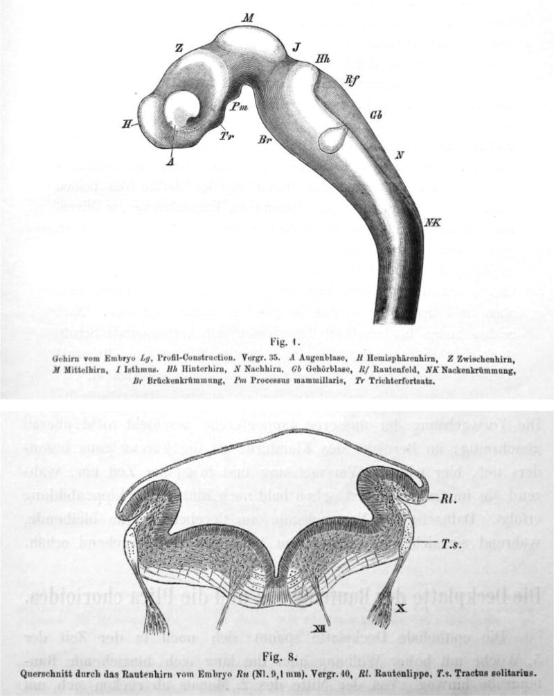
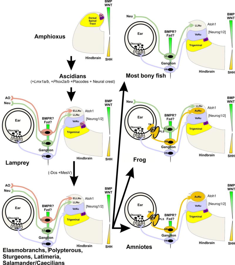
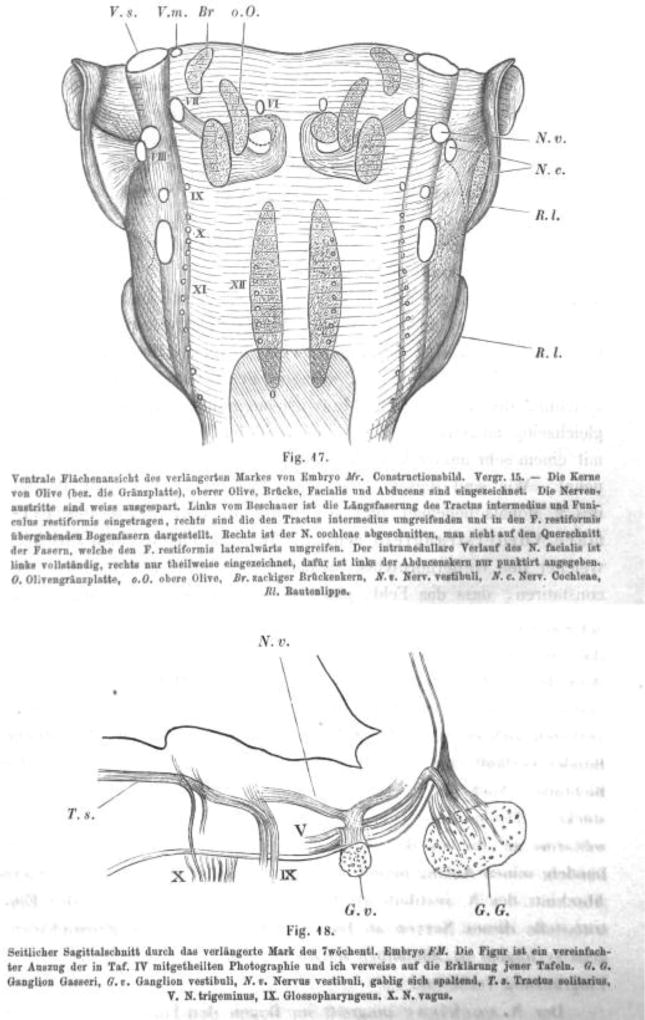
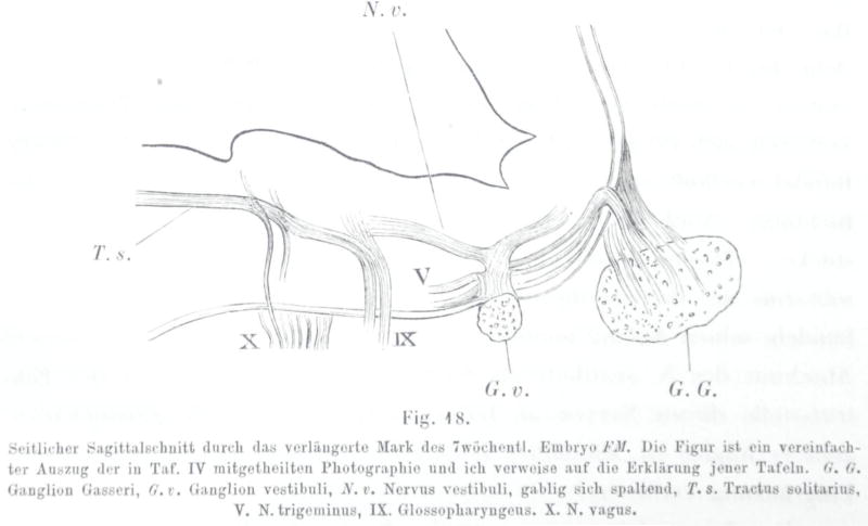
Wilhelm His (1831-1904) provided lasting insights into the development of the central and peripheral nervous system using innovative technologies such as the microtome, which he invented. 150 years after his resurrection of the classical germ layer theory of Wolff, von Baer and Remak, his description of the developmental origin of cranial and spinal ganglia from a distinct cell population, now known as the neural crest, has stood the test of time and more recently sparked tremendous advances regarding the molecular development of these important cells. In addition to his 1868 treatise on 'Zwischenstrang' (now neural crest), his work on the development of the human hindbrain published in 1890 provided novel ideas that more than 100 years later form the basis for penetrating molecular investigations of the regionalization of the hindbrain neural tube and of the migration and differentiation of its constituent neuron populations. In the first part of this review we briefly summarize the major discoveries of Wilhelm His and his impact on the field of embryology. In the second part we relate His' observations to current knowledge about the molecular underpinnings of hindbrain development and evolution. We conclude with the proposition, present already in rudimentary form in the writings of His, that a primordial spinal cord-like organization has been molecularly supplemented to generate hindbrain 'neomorphs' such as the cerebellum and the auditory and vestibular nuclei and their associated afferents and sensory organs.


Figures

Fig 1
Fig 1
(Showing His, 1890 Fig.1) Brain of embryo Lg, profile-construction, Mag 35, a, eye vesicle, H, forebrain hemisphere, Z, diencephalon; M, mesencephalon; I, isthmus; Hh, pons; N, medulla oblongata; Gb, ear vesicle; Rf, rhombencephalon; NK, spinal flexure; Br, pontine flexure; Pm, mammillary bodies; Tr; pituitary. Note the shaded area below RF that shows the choroid plexus (not labeled as such by His).
Fig. 2
Fig. 2
(Showing His, 1890 Fig. 8) Coronal section through the rhombencephalon of embryo Bm, Nl 0.1 mm, Mag 40, Rl rhombic lip; Ts, solitary tract.
Fig 3
Fig 3
(Showing His, 1890; Fig. 17) Ventral view of the rhombencephalon of embryo Mr, image of a reconstruction, Ma 15 – The olivary nuclei (or pons), superior olive, pons, facial, abducens are shown). Nerve roots are shown in white. The left shows the intermediate tract and the restiform body, the right shows the arcuate fibers surrounding the intermediate and pass to the restiform body. The right cochlear nucleus is removed to show the fibers that surround laterally the restiform body. The intramedullary course of the facial nerve is completely shown on the left, but only partially on the right, but the abducens is only outlined by a dotted line. G, pons, oO superior olive, Br, dentate potine nucleus; Nv, vestibular nerve; Nc, cochlear nerve; Rl, rhombic lip.
Fig. 4
Fig. 4
(Showing His, 1890; Fig. 18) Parasagittal section of the rhombencephalon of the 7 week old embryo FM. This Fig is a simplified excerpt of the photograph in plate IV and I refer to this plate for explanations. Gg, Gasserian ganglion, Gv vestibular ganglion, Nv, vestibular nerve, bifurcating; Ts, solitary tract; V, descending tract of V; IX Glossopharyngeus; X, vagus.
Fig. 5
Fig. 5
The evolution of gene expression at the midbrain–hindbrain boundary (MHB) is shown for deuterostomes. The MHB of vertebrates exhibits abutting domains of Otx2 and Gbx2 expression (d–g). This stabilizes the expression of Fgf8 (g), which in turn stabilizes the expression of Wnt1 and engrailed (En1). Mutation of Otx2, Gbx2, Fgf8, or Wnt1 eliminates the MHB. Pax2/5/8 are also expressed at the MHB, whereas the expression of Dmbx occurs immediately rostral to the MHB in the midbrain to later expand into the hindbrain and spinal cord (d). Note the partial overlap of Pax2/5/8 with the caudal expression of Otx2 and the rostral expression of Gbx2 (d). Hemichordates (a) have overlapping expression of Gbx, Otx, Irx and En in the rostral trunk. Pax6 abuts Gbx2 whereas Pax2/5/8 overlaps with the caudal expression of Gbx2. Outgroup data suggest that coelenterates have a Dmbx ortholog, thus raising the possibility that hemichordates (a) also have a Dmbx gene. Cephalochordates (b) have no Dmbx expression in the ‘brain’. The Otx expression domain abuts the Gbx expression domain, as in vertebrates. However, Gbx overlaps with Pax2/5/8 and most of Irx3. Urochordates (c) have no Gbx gene but have a Pax2/5/8 and Pax6 configuration comparable to vertebrates. Dmbx overlaps with the caudal end of the Irx3 expression whereas Dmbx expression is rostral to Irx3 in vertebrates. Together, these data show that certain gene expression domains are topographically conserved (Foxg1, Hox, Otx), whereas others show varying degrees of overlap. It is conceivable that the evolution of nested expression domains of transcription factors is causally related to the evolution of specific neuronal features such as the evolution of oculomotor and trochlear motoneurons (d, e) around the MHB. Experimental work has demonstrated that the development of these motor centers depends on the formation of the MHB. Modified after (Fritzsch and Glover 2006; Guo et al. 2007; Mishima et al. 2009; Fritzsch et al. 2015; Albuixech-Crespo et al. 2017))
Fig. 6
Fig. 6
Loss of Lmx1a/b results in absence of the typical rhombencephalic form with a IVth ventricle covered by a choroid plexus. It also eliminates the midbrain-hindbrain boundary and thus anterior hindbrain/midbrain structures such as oculomotor and trochlear motneurons and the substantia nigra. The cross sections of the alar plate show the partially overlapping expression of different factors in the alar plate that regulate development of subpopulations in rhombomere 7. Note that the expression domains of bHLH genes in r1–5 are only partially clarified. Note also that loss of Lmx1a/b eliminates expression of Atoh1 and Wnt1 throughout most of the hindbrain. Modified after (Mishima et al. 2009; Ray and Dymecki 2009)
Fig. 7
Fig. 7. Phylogenetic analysis of the chordate LMX1 protein family
Vertebrate LMX1A and LMX1B subclades are depicted in red and blue, respectively, and the predicted LMX1 peptide from the Japanese lamprey (L. japonicum JL14965) is in purple (REF #1). Non-vertebrate, chordate out-group sequences (black) consist of a pair of duplicated LMX1 genes, lmx-like and lmx, which are present in the genomes of each of the two ascidians Ciona intestinalis and Ciona savignyi. Node values represent posterior probabilities after phylogenetic analysis via Bayesian inference.
Fig. 8
Fig. 8. Proposed octavo-lateral evolution
Evolution from the common ancestor of Amphioxus and Lamprey resulted in the eventual expansion of the alar plate to accommodate the vestibular, lateral line, and electroreceptive nuclei. Sharks, Latimeria, and salamanders have lost Dorsal cells (DC), but acquired the sensory MesV nucleus. Most bony fish have retained a lateral line projection, but have lost electroreception. Similarly, frogs have done the same, but have additionally gained auditory nuclei. Amniotes have lost both lateral line projections and electroreception, but have retained auditory nuclei. AO, ampullary organs; Neu, neuromasts; ST, solitary tract. Modified after (Fritzsch and Elliott 2017)

Similar articles

Cited by

References

    1. Albuixech-Crespo B, López-Blanch L, Burguera D, Maeso I, Sánchez-Arrones L, et al. Molecular regionalization of the developing amphioxus neural tube challenges major partitions of the vertebrate brain. PLoS biology. 2017;15:e2001573. - PMC - PubMed
    1. Atwell WJ, Danchakoff V. A human embryo with seventeen pairs of somites. Carnegie Institution of Washington; 1930.
    1. Bermingham NA, Hassan BA, Wang VY, Fernandez M, Banfi S, et al. Proprioceptor pathway development is dependent on Math1. Neuron. 2001;30:411–422. - PubMed
    1. Bonito M, Glover JC, Studer M. Hox genes and region-specific sensorimotor circuit formation in the hindbrain and spinal cord. Developmental Dynamics. 2013;242:1348–1368. - PubMed
    1. Burzynski GM, Reed X, Maragh S, Matsui T, McCallion AS. Integration of genomic and functional approaches reveals enhancers at LMX1A and LMX1B. Molecular genetics and genomics. 2013;288:579–589. - PMC - PubMed

Personal name as subject

LinkOut - more resources