

Progression of Primary Open-Angle Glaucoma in Diabetic and Nondiabetic Patients
- PMID: 29447914
- PMCID: PMC5916320
- DOI: 10.1016/j.ajo.2018.02.002
Progression of Primary Open-Angle Glaucoma in Diabetic and Nondiabetic Patients
Abstract
Purpose: To compare the rates of visual field (VF) loss and retinal nerve fiber layer (RNFL) thinning in primary open-angle glaucoma (POAG) patients with or without type 2 diabetes mellitus (DM).
Design: Cohort study.
Methods: A total of 197 eyes (55 eyes of 32 POAG patients with DM in POAG/DM group and 142 eyes of 111 age-matched POAG patients without DM in POAG/DM- group) were included from the Diagnostic Innovations in Glaucoma Study (DIGS). Type 2 DM participants were defined by self-report of DM history and use of antidiabetic medication. The rates of VF loss and RNFL loss were compared in POAG eyes with and without DM using univariate and multivariable mixed-effects models.
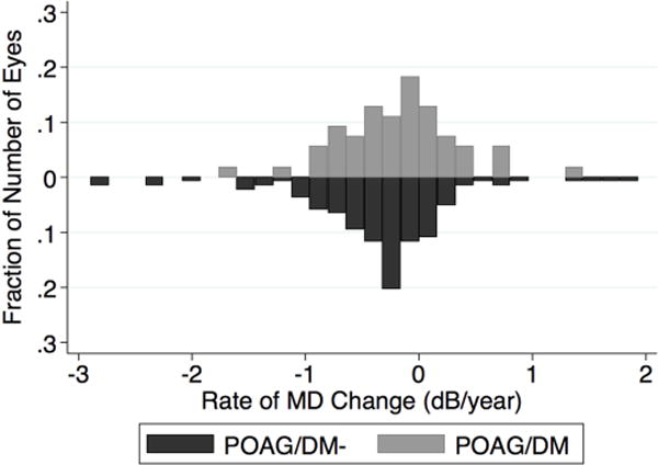
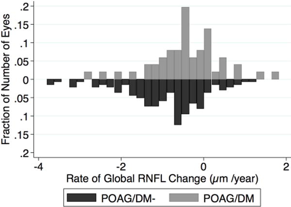
Results: The median (interquartile range) follow-up was 5.7 years (4.0, 6.4). The mean rate of global RNFL loss in the POAG/DM group was 2-fold slower than in the POAG/DM- group overall (-0.40 μm/year vs -0.83 μm/year, respectively P = .01). Although a slower rate of VF mean deviation and pattern standard deviation loss was found in the POAG/DM group compared to the POAG/DM- group, the difference was not statistically significant.
Conclusions: POAG patients with treated type 2 DM, who had no detectable diabetic retinopathy, had significantly slower rates of RNFL thinning compared to those without diagnosed DM.
Copyright © 2018 Elsevier Inc. All rights reserved.
Figures
References
-
- Weinreb RN, Khaw PT. Primary open-angle glaucoma. Lancet. 2004;363:1711–20. - PubMed
-
- Tham YC, Cheng CY. Associations between chronic systemic diseases and primary open angle glaucoma: an epidemiological perspective. Clin Exp Ophthalmol. 2017;45:24–32. - PubMed
-
- Gordon MO, Beiser JA, Brandt JD, et al. The Ocular Hypertension Treatment Study: baseline factors that predict the onset of primary open-angle glaucoma. Arch Ophthalmol. 2002;120:714–20. discussion 829–30. - PubMed
Publication types
MeSH terms
Grants and funding
LinkOut - more resources
Full Text Sources
Other Literature Sources
Medical
