. 2018 Apr 15:1685:51-59.
doi: 10.1016/j.brainres.2018.02.005. Epub 2018 Feb 12.

Intrauterine inflammation reduces postnatal neurogenesis in the hippocampal subgranular zone and leads to accumulation of hilar ectopic granule cells

Affiliations

Intrauterine inflammation reduces postnatal neurogenesis in the hippocampal subgranular zone and leads to accumulation of hilar ectopic granule cells

Michael S Hester et al. Brain Res. .

Abstract

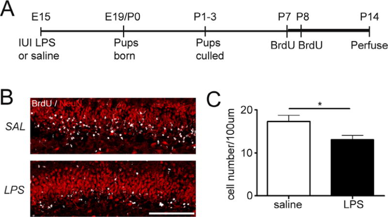
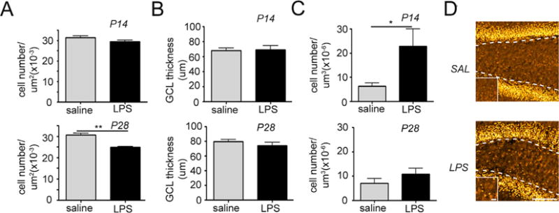
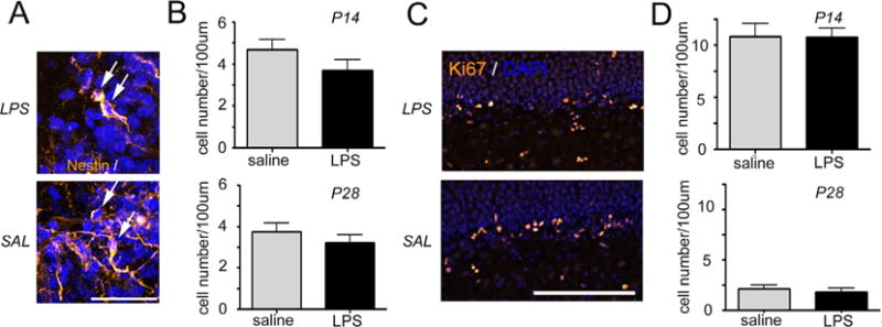
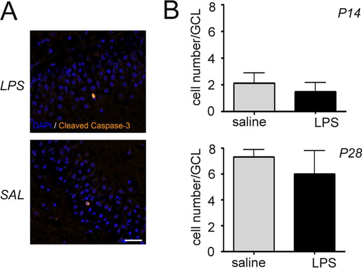
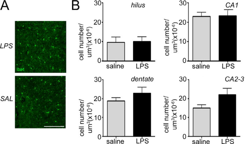
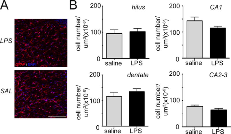
Prenatal inflammation is associated with poor neurobehavioral outcomes in exposed offspring. A common route of exposure for the fetus is intrauterine infection, which is often associated with preterm birth. Hippocampal development may be particularly vulnerable to an inflammatory insult during pregnancy as this region remains highly neurogenic both prenatally and postnatally. These studies sought to determine if intrauterine inflammation specifically altered hippocampal neurogenesis and migration of newly produced granule neurons during the early postnatal period. Microglial and astroglial cell populations known to play a role in the regulation of postnatal neurogenesis were also examined. We show that intrauterine inflammation significantly reduced hippocampal neurogenesis between postnatal days 7 (P7) and P14 as well as decreased granule cell density at P28. Ectopic migration of granule cells was observed in LPS-exposed mice at P14, but not at P28. Intrauterine inflammation had no effect on hippocampal astrocyte or microglia density or on apoptosis rate at the postnatal time points examined. Thus, exposure to intrauterine inflammation disrupts early postnatal neurogenesis and leads to aberrant migration of newly born granule cells.

Keywords: Astroglia; Ectopic granule cells; Fetal brain injury; Hippocampal neurogenesis; Intrauterine inflammation; Microglia.


Conflict of interest statement

Additional information

The authors declare no conflict of interest.

Figures

Fig. 1
Fig. 1
Exposure to intrauterine inflammation reduces early postnatal neurogenesis. (A) Schematic of experimental design. Pregnant dams were given intrauterine injections of lipopolysaccharide (LPS) [50 μg] or saline on embryonic day 15. Pups delivered at term were culled [5 pups/dam] and given injections of bromodeoxyuridine [BrdU, 75 mg/kg; s.c.] at postnatal days 7 and 8 (P7–P8) to label actively proliferating cells. Mice were transcardially perfused with paraformaldehyde at postnatal day 14 (B). Confocal maximum projection images of BrdU (white) and Neuronal Nuclei (NeuN, red) co-labeling in the dentate gyrus of mice exposed to intrauterine saline or LPS and sacrificed at P14. Scale bar = 100 μm. (C) Quantification of BrdU/NeuN density in the granule cell layer at P14. LPS: n = 10; saline: n = 8. *p<0.05. Data represent the mean ± SEM.
Fig. 2
Fig. 2
Intrauterine inflammation leads to decreased total neuronal density and transient migration abnormalities in the dentate gyrus. Quantification of granule cell density in the granule cell layer (GCL) (A), granule cell layer thickness (B), and ectopic granule cell density in the hilus (C) at postnatal days 14 and 28. *p < 0.05. Data represent the mean ± SEM. (D) Confocal maximum projection images of the granule cell-specific marker Prospero Homeobox 1 (Prox1) in the dentate gyrus of mice exposed to intrauterine saline or lypopolysaccharide (LPS) and sacrificed at P14 (LPS: n = 10; saline: n = 8). Dotted lines depict the granule cell layer (GCL)/hilar border. High magnification images depicted on the bottom left were taken from the center of the hilus. Scale bar = 100 μm, magnified panel = 10 μm.
Fig. 3
Fig. 3
Intrauterine inflammation has no effect on the number and division rate of neuronal progenitor cells. Confocal maximum projection images of Nestin (A, yellow) and Ki67 (C, yellow) labeling of the neural progenitor cells in the dentate gyrus of mice exposed to intrauterine saline or lipopolysaccharide (LPS) and sacrificed at postnatal day 14 (P14). Arrows depict Nestin-positive neuronal progenitor cells in the subgranular zone (A). Cell nuclei are counterstained with 4′,6-diamidino-2-phenylindole (DAPI, blue). Scale bars are 20 μm (A) and 100 μm (C). Quantification of neuronal progenitor cell density (B) and the density of the Ki67 labeled cells (D) in the subgranular zone at P14 and P28. Data represent the mean ± SEM. P14: LPS, n = 10; saline, n = 8; P28: LPS n = 6; saline n = 6.
Fig. 4
Fig. 4
Intrauterine inflammation has no effect on postnatal apoptosis in the dentate gyrus. (A) Confocal maximum projection images through the dentate gyrus of mice exposed to intrauterine saline or lipopolysaccharide (LPS) and sacrificed at postnatal day 14 (P14). Apoptotic cells are labeled with antibodies against Cleaved Caspase-3 (yellow). 4′,6-diamidino-2-phenylindole (DAPI, blue) labels cell nuclei. Scale bar = 20 μm. (B) Quantification of Cleaved Caspase-3-expressing cells in the granule cell layer of the dentate gyrus at P14 and P28. Data represent the mean ± SEM. P14: LPS, n = 10; saline, n = 8; P28: LPS n = 6; saline n = 6.
Fig. 5
Fig. 5
Intrauterine inflammation has no effect on hippocampal microglia density. (A) Confocal maximum projection images of the microglia-specific marker Ionized calcium-binding adapter molecule 1 (Iba1, green) in the Cornu Ammonis 1 (CA1) hippocampal region of mice exposed to intrauterine saline or lipopolysaccharide (LPS) and sacrificed at postnatal day 28 (P28). 4′,6-diamidino-2-phenylindole (DAPI) counterstains cell nuclei (blue). Scale bar = 100 μm. (B) Quantification of microglia density in the dentate gyrus, hilus, CA1, and CA2-3 regions at P14. Data represent the mean ± SEM. P14: LPS, n = 10; saline, n = 8; P28: LPS n = 6; saline n = 6.
Fig. 6
Fig. 6
Intrauterine inflammation has no effect on hippocampal astrocyte density. (A) Confocal maximum projection images of the astrocyte-specific marker Glial Fibrillary Acidic Protein (GFAP, red) in the Cornu Ammonis 1 (CA1) hippocampal region of mice exposed to intrauterine saline or lipopolysaccharide (LPS) and sacrificed at postnatal day (P14). Cell nuclei are labeled with 4′,6-diamidino-2-phenylindole (DAPI) counterstain (blue). Scale bar = 100 μm. (B) Quantification of astrocyte density in the dentate gyrus, hilus, CA1, and CA2-3 regions at P14. Data represent the mean ± SEM. P14: LPS, n = 10; saline, n = 8.

References

    1. Aguilar-Valles A, Luheshi GN. Alterations in cognitive function and behavioral response to amphetamine induced by prenatal inflammation are dependent on the stage of pregnancy. Psychoneuroendocrinology. 2011;36:634–648. - PubMed
    1. Aimone JB, Li Y, Lee SW, Clemenson GD, Deng W, Gage FH. Regulation and function of adult neurogenesis: from genes to cognition. Physiol Rev. 2014;94:991–1026. - PMC - PubMed
    1. Allen MC. Neurodevelopmental outcomes of preterm infants. Curr Opin Neurol. 2008;21:123–128. - PubMed
    1. Ambrogini P, Lattanzi D, Ciuffoli S, Agostini D, Bertini L, Stocchi V, Santi S, Cuppini R. Morpho-functional characterization of neuronal cells at different stages of maturation in granule cell layer of adult rat dentate gyrus. Brain Res. 2004;1017:21–31. - PubMed
    1. Anderson P, Doyle LW. Neurobehavioral outcomes of school-age children born extremely low birth weight or very preterm in the 1990s. JAMA. 2003;289:3264–3272. - PubMed

Publication types

MeSH terms

Substances