. 2018 Feb 15;6(1):9.
doi: 10.1186/s40478-018-0510-8.

Brain region-specific enhancement of remyelination and prevention of demyelination by the CSF1R kinase inhibitor BLZ945

Affiliations

Brain region-specific enhancement of remyelination and prevention of demyelination by the CSF1R kinase inhibitor BLZ945

Nicolau Beckmann et al. Acta Neuropathol Commun. .

Abstract

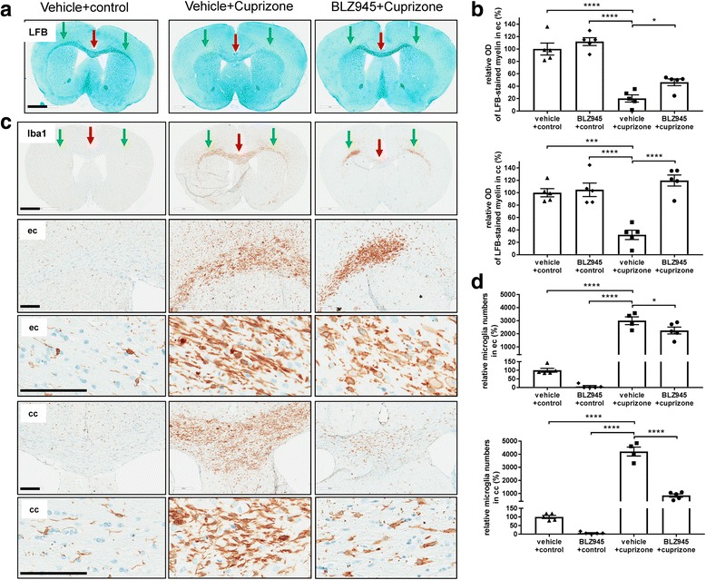
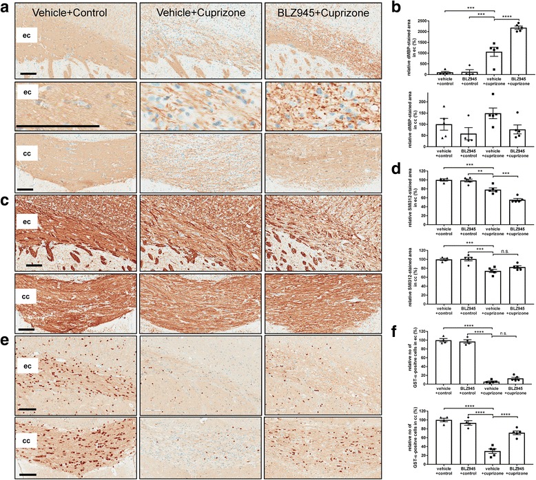
Multiple sclerosis (MS) is a chronic inflammatory disease affecting the central nervous system (CNS). While multiple effective immunomodulatory therapies for MS exist today, they lack the scope of promoting CNS repair, in particular remyelination. Microglia play a pivotal role in regulating myelination processes, and the colony-stimulating factor 1 (CSF-1) pathway is a key regulator for microglia differentiation and survival. Here, we investigated the effects of the CSF-1 receptor kinase inhibitor, BLZ945, on central myelination processes in the 5-week murine cuprizone model by non-invasive and longitudinal magnetic resonance imaging (MRI) and histology. Therapeutic 2-week BLZ945 treatment caused a brain region-specific enhancement of remyelination in the striatum/cortex, which was absent in the corpus callosum/external capsule. This beneficial effect correlated positively with microglia reduction, increased oligodendrocytes and astrogliosis. Prophylactic BLZ945 treatment prevented excessive demyelination in the corpus callosum by reducing microglia and increasing oligondendrocytes. In the external capsule oligodendrocytes were depleted but not microglia and a buildup of myelin debris and axonal damage was observed. A similar microglial dysfunction in the external capsule with an increase of myelin debris was obvious in triggering receptor expressed on myeloid cells 2 (TREM2) knock-out mice treated with cuprizone. Finally, therapeutic BLZ945 treatment did not change the disease course in experimental autoimmune encephalomyelitis mice, a peripherally driven neuroinflammation model. Taken together, our data suggest that a short-term therapeutic inhibition of the CSF-1 receptor pathway by BLZ945 in the murine cuprizone model enhances central remyelination by modulating neuroinflammation. Thus, microglia-modulating therapies could be considered clinically for promoting myelination in combination with standard-of-care treatments in MS patients.

Keywords: Astrocyte; BLZ945; CSF1R kinase; Cuprizone; Microglia; Multiple sclerosis; Myelination; Neuroinflammation; Oligodendrocyte; TREM2.


Conflict of interest statement

Competing interests

All authors are, or have been, employees and shareholders of Novartis Pharma AG, Basel Switzerland.

Publisher’s Note

Springer Nature remains neutral with regard to jurisdictional claims in published maps and institutional affiliations.

Figures

Fig. 1
Fig. 1
MRI reliably detects de- and re-myelination events in the cuprizone model in the corpus callosum/external capsule, correlating with myelin and oligodendrocyte histology. a Representative MRI images acquired from two mice, one receiving normal food (left) and the other treated with 0.2% cuprizone for 5 weeks with subsequent switch to normal food. b Representative pictures from histological and immunohistological stainings from the corpus callosum/external capsule after 5-week 0.2% cuprizone food and control food treatment and recovery for 2 and 4 weeks (switch to normal food) of C57BL/6 mice detecting myelin by Luxol fast blue (LFB), myelin oligondendrocyte glycoprotein (MOG) and GST-π positive oligodendrocyte cells in the corpus callosum/external capsule. c Correlation analysis of quantitative histology (LFB and MOG optical density (OD), GST-π positive soma area normalized (norm.) to region of interest) and MRI signal/MTR parameters. Two-tailed Pearson correlation analysis, correlation coefficients (R2) and p values are indicated. Scale bars: 400 μm. cc: corpus callosum, ec: external capsule, MRI: magnetic resonance imaging, MTR: magnetization transfer ratio, a.u.: arbitrary units, OD: optical density
Fig. 2
Fig. 2
A 2-week therapeutic treatment with BLZ945 after a 5-week cuprizone intoxication period reduced MRI signal in cortex and striatum but not corpus callosum, suggesting increased remyelination. a Schematic diagram of the experimental setup for the therapeutic treatment. Groups consisted of mice treated for 5 weeks with control food (normal food) or 0.2% cuprizone in food and then switched back to control food (normal food) for the 2-week therapeutic treatment (vehicle or BLZ945 169 mg/kg p.o., qd). MRI measurements were performed at week 0 (baseline), week 5 at max. Pathology of cuprizone intoxication, at week 6 (1 week of therapeutic or vehicle treatment on control food) and at week 7 (2 weeks of therapeutic or vehicle treatment on control food). Mice were killed at week 7 immediately after the last MRI measurement. b Representative MRI images acquired from two mice, one receiving 0.2% cuprizone and then normal food with vehicle (upper row) and the other treated with 0.2% cuprizone for 5 weeks with subsequent switch to normal food and BLZ945 treatment (lower row). c Representative MRI images indicating analyzed brain regions (in red). d MRI signal in cortex and striatum for the different treatment groups. For each brain region, MRI signal was normalized to absolute values in the control group (control food, vehicle treatment). e MRI signal and MTR in corpus callosum and external capsule for the different treatment groups (normalized to values in the control group). Because of the small magnitude (≤2%) of MTR reductions in the cortex and striatum following the 5-week cuprizone intoxication period, MTR changes in these areas were not considered here. Grey and black symbols indicate individual values from two independent experiments. Group sizes: control+vehicle (n = 7 from experiment 1, n = 7 from experiment 2), cuprizone+vehicle (n = 6 from experiment 1, n = 6 from experiment 2; one mouse was removed from experiment 2 due to technical reasons), cuprizone+BLZ945 (n = 7 from experiment 1, n = 6 from experiment 2). Data is shown as mean ± SEM. Statistics (for combined experiments): Turkey’s multiple comparison test (***: p < 0.001, ****: p < 0.0001), n.s.: not significant, ctrl: control, cpz: cuprizone, cc: corpus callosum, ec: external capsule, MRI: magenetic resonance imaging, MTR: magnetization transfer ratio
Fig. 3
Fig. 3
A 2-week therapeutic treatment with BLZ945 after a 5-week cuprizone intoxication period enhanced remyelination and increased the number of mature oligodendrocytes in cortex and striatum but not corpus callosum/external capsule compared to vehicle treatment. a Representative pictures from immunohistological stainings detecting myelin basic protein (MBP) and mature oligodendrocytes positive for GST-π in the cortex for the different treatment groups at week 7 (see Fig. 2a for the experimental setups and groups). b, c Corresponding quantitative analysis of the immunohistochemistry for MBP (stained area) and GST-π (number of positive cells) in the cortex and striatum normalized to values from control vehicle mice. d Representative pictures from immunohistological stainings detecting myelin oligondendrocyte glycoprotein (MOG) and mature oligodendrocytes positive for GST-π in the corpus callosum and external capsule for the different treatment groups at week 7. e Corresponding analysis of the immunohistochemistry for MOG (optical density, OD) and GST-π (number of positive cells) as well as OD analysis of Luxol fast blue (LFB) in the cc and ec. Values were normalized to those of control vehicle mice. Grey and black symbols indicate individual values from two independent experiments. Group sizes: control+vehicle (n = 7 from experiment 1, n = 6–7 from experiment 2), cuprizone+vehicle (n = 7 from experiment 1, n = 7 from experiment 2), cuprizone+BLZ945 (n = 7 from experiment 1, n = 5–6 from experiment 2). Data are shown as means±SEM. Scale bars: 200 μm (MBP and MOG), 100 μm (GST-π). Statistics (for combined experiments): Turkey’s multiple comparison test one-way ANOVA (*: p < 0.05, **: p < 0.01, ***: p < 0.001, ****: p < 0.0001, n.s.: not significant), cpz: cuprizone, cc: corpus callosum, ec: external capsule, OD: optical density
Fig. 4
Fig. 4
A 2-week therapeutic treatment with BLZ945 after a 5-week cuprizone intoxication period reduced microglia numbers but enhanced astrocytes. a Representative pictures from immunohistological stainings detecting the microglia marker Iba1 and glial fibrillary acidic protein (GFAP) astrocytes in the cortex for the different treatment groups at week 7 (see Fig. 2a for the experimental setup and groups). b, c, d Corresponding quantitative analysis of the immunohistochemistry for Iba1-positive microglia numbers and GFAP-positive astrocyte stained area in the cortex, striatum and corpus callosum/external capsule. Values were normalized to those of control, vehicle-treated mice. Group sizes: For all treatments n = 7. Data are shown as means±SEM. Scale bars: 100 μm. Statistics: Turkey’s multiple comparison test one-way ANOVA (**: p < 0.01, ***: p < 0.001, ****: p < 0.0001, n.s.: not significant), cpz: cuprizone, cc: corpus callosum, ec: external capsule
Fig. 5
Fig. 5
Prophylactic treatment with BLZ945 1 week before and during 5-week cuprizone intoxication inhibited demyelination in the corpus callosum but not in the external capsule. a Schematic diagram of the experimental setup for the prophylactic treatment. Groups consisted of mice pretreated for 1 week with vehicle or 169 mg/kg BLZ945 (p.o., qd) on normal food. Control food was continued or then switched to 0.2% cuprizone with concomitant continuation of vehicle or 169 mg/kg BLZ945 (p.o., qd) treatment for 5 weeks. MRI measurements were performed at week 0 (baseline) as well as week 3 and week 5 during cuprizone intoxification. Mice were killed at week 5 immediately after the last MRI measurement. b Representative MRI brain images for mice treated with either BLZ945 (p.o., qd, 169 mg/kg) or vehicle before (1 week) and during 0.2% cuprizone intoxification for 5 weeks. red arrows: corpus callosum, green arrows: external capsule. c Representative MRI brain images indicating the analyzed brain regions (red: corpus callosum, green: external capsule). d, e Quantification of the MRI contrast and MTR in the corpus callosum for the different treatment groups showing reduced MRI contrast and enhanced MTR for the corpus callosum compared to the external capsule. For all groups: n = 5. Data is shown as mean ± SEM. Statistics: Turkey’s multiple comparison test one-way ANOVA (*: p < 0.05, **: p < 0.01, ***: p < 0.001, ****: p < 0.0001), n.s.: not significant, a.u.: arbitrary units, MRI: magnetic resonance imaging, MTR: magnetization transfer ratio
Fig. 6
Fig. 6
Prophylactic treatment with BLZ945 1 week before and during 5-week cuprizone intoxication inhibited demyelination and reduced microglia in the corpus callosum but enhanced axonal pathology and myelin debris in the external capsule. a Representative overview pictures from histological stainings of Luxol Fast Blue (LFB) for the different treatment groups at week5 (see Fig. 5a for the experimental setup and groups), red arrows: corpus callosum, green arrows: external capsule. b Corresponding analysis of the optical density (OD) of Luxol fast blue (LFB) in the cc and ec. c Representative overview and higher magnification pictures from immunohistological stainings detecting Iba1-positive microglia for the different treatment groups at week5 (see Fig. 5a for the experimental setup and groups), red arrows: corpus callosum, green arrows: external capsule. d Corresponding quantitative analysis of the immunohistochemistry for Iba1-positive microglia numbers in the corpus callosum and external capsule. Values were normalized to those of control vehicle mice. Group sizes: for all treatment groups n = 4–5. Data are shown as means±SEM. Scale bars: 200 μm for the higher magnification. Statistics: Turkey’s multiple comparison test one-way ANOVA (*: p < 0.05, ***: p < 0.001, ****: p < 0.0001), cpz: cuprizone, cc: corpus callosum, ec: external capsule, OD: optical density
Fig. 7
Fig. 7
Prophylactic treatment with BLZ945 1 week before and during 5-week cuprizone intoxication revealed reduced oligodendrocyte numbers, appearance of myelin debris and axonal pathology in the external capsule but not in the corpus callosum. a Representative images from immunohistological stainings in the external capsule and corpus callosum detecting debris of myelin basic protein (dMBP). Myelin debris were obvious in the external capsule with BLZ945 and cuprizone treatment while no obvious change could be observed in the corpus callosum. b Corresponding quantitative analysis of the immunohistochemistry for dMBP-positive area in the corpus callosum and external capsule. Please note: The threshold for the staining of the control groups were below the image analysis parameters used. Values were normalized to those of control vehicle mice. c Representative images from immunohistological stainings in the external capsule and corpus callosum detecting neurofilament (SMI312) for the different treatment groups at week 5. Loss of axonal neurofilaments was obvious in the external capsule with BLZ945 and cuprizone treatment while no change could be observed in the corpus callosum. d Corresponding quantitative analysis of the immunohistochemistry for SMI312-positive area in the corpus callosum and external capsule. e Representative images from immunohistological stainings in the external capsule and corpus callosum detecting mature oligodendrocytes (GST-π) for the different treatment groups at week 5. Reduced oligodendrocyte numbers were observed in the external capsule while an increase in the corpus callosum was obvious with BLZ945 and cuprizone treatment. f Corresponding quantitative analysis of the immunohistochemistry for GST-π-positive soma numbers in the corpus callosum and external capsule. Values were normalized to those of control vehicle mice. Group sizes: for all treatment groups n = 4–5. Data are shown as means±SEM. Scale bars: 100 μm. Statistics: Turkey’s multiple comparison test one-way ANOVA **: p < 0.01, ***: p < 0.001, ****: p < 0.0001, n.s.: not significant), cpz: cuprizone, cc: corpus callosum, ec: external capsule

References

    1. Asai H, Ikezu S, Tsunoda S, Medalla M, Luebke J, Haydar T, Wolozin B, Butovsky O, Kügler S, Ikezu T. Depletion of microglia and inhibition of exosome synthesis halt tau propagation. Nat Neurosci. 2015;18:1584–1593. doi: 10.1038/nn.4132. - DOI - PMC - PubMed
    1. Brück W, Pförtner R, Pham T, Zhang J, Hayardeny L, Piryatinsky V, Hanisch U-K, Regen T, van Rossum D, Brakelmann L, Hagemeier K, Kuhlmann T, Stadelmann C, John GR, Kramann N, Wegner C. Reduced astrocytic NF-κB activation by laquinimod protects from cuprizone-induced demyelination. Acta Neuropathol. 2012;124:411–424. doi: 10.1007/s00401-012-1009-1. - DOI - PMC - PubMed
    1. Calabrese M, Magliozzi R, Ciccarelli O, Geurts JJG, Reynolds R, Martin R. Exploring the origins of grey matter damage in multiple sclerosis. Nat Rev Neurosci. 2015;16:147–158. doi: 10.1038/nrn3900. - DOI - PubMed
    1. Cantoni C, Bollman B, Licastro D, Xie M, Mikesell R, Schmidt R, Yuede CM, Galimberti D, Olivecrona G, Klein RS, Cross AH, Otero K, Piccio L (2015) TREM2 regulates microglial cell activation in response to demyelination in vivo. Acta Neuropathol. doi: 10.1007/s00401-015-1388-1 - PMC - PubMed
    1. Chandran P, Upadhyay J, Markosyan S, Lisowski a, Buck W, Chin C-L, Fox G, Luo F, Day M. Magnetic resonance imaging and histological evidence for the blockade of cuprizone-induced demyelination in C57BL/6 mice. Neuroscience. 2012;202:446–453. doi: 10.1016/j.neuroscience.2011.10.051. - DOI - PubMed

MeSH terms

Substances