Review
. 2018 Feb 15;16(1):29.
doi: 10.1186/s12967-018-1402-1.

Non-viral gene delivery systems for tissue repair and regeneration

Affiliations
Review

Non-viral gene delivery systems for tissue repair and regeneration

Pan Wu et al. J Transl Med. .

Abstract

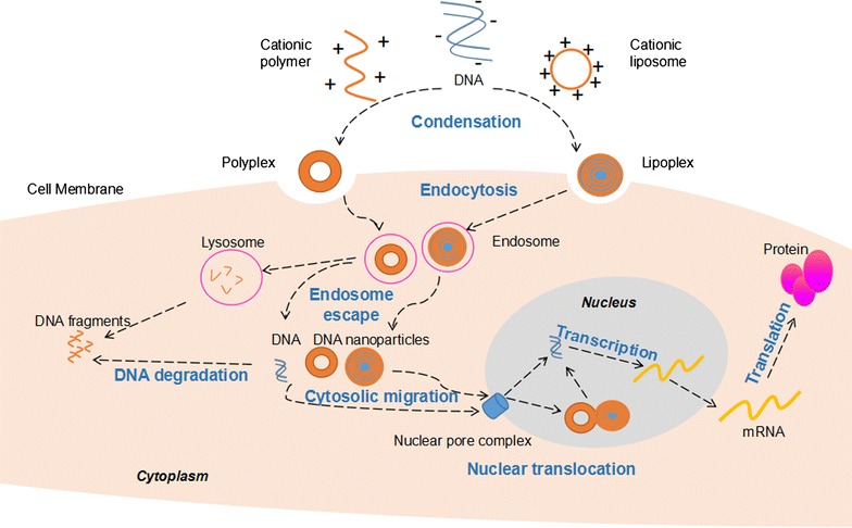
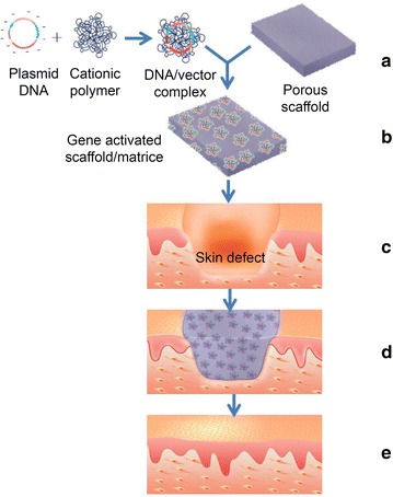
Critical tissue defects frequently result from trauma, burns, chronic wounds and/or surgery. The ideal treatment for such tissue loss is autografting, but donor sites are often limited. Tissue engineering (TE) is an inspiring alternative for tissue repair and regeneration (TRR). One of the current state-of-the-art methods for TRR is gene therapy. Non-viral gene delivery systems (nVGDS) have great potential for TE and have several advantages over viral delivery including lower immunogenicity and toxicity, better cell specificity, better modifiability, and higher productivity. However, there is no ideal nVGDS for TRR, hence, there is widespread research to improve their properties. This review introduces the basic principles and key aspects of commonly-used nVGDSs. We focus on recent advances in their applications, current challenges, and future directions.

Keywords: Gene therapy; Non-viral vector; Tissue engineering.


Figures

Fig. 1
Fig. 1
Basic mechanism of non-viral gene delivery via polyplex and lipoplex. DNA is condensed via interaction with a cationic polymer or encapsulated in a cationic liposome to form a polyplex or lipoplex and pass through the cell membrane via endocytosis. Once endosome escape occurs, the complex is released into the cytosol, the released DNA will be transported to the perinuclear region via microtubule system. Nuclear translocation of the exogenous DNA can be achieved by passage through nuclear pore complex in non-dividing cells. After right transcription and translation, target proteins are produced to exert biological effects
Fig. 2
Fig. 2
Schematic illustrating the construction of a gene activated scaffold/matrice and its application in skin defect. (a) Formation of a plasmid DNA/cationic polymer complex which is then loaded onto a scaffold. (b) A gene activated scaffold/matrice. (c) A deep skin defect. (d) Transplantation of a gene activated scaffold/matrice which fills the skin defect. (e) vascularization of the scaffold accompanied with repair and regeneration of the skin

References

    1. Peck MD. Epidemiology of burns throughout the world. Part I: distribution and risk factors. Burns. 2011;37(7):1087–1100. doi: 10.1016/j.burns.2011.06.005. - DOI - PubMed
    1. Dimitriou R, Jones E, McGonagle D, Giannoudis PV. Bone regeneration: current concepts and future directions. BMC Med. 2011;9:66. doi: 10.1186/1741-7015-9-66. - DOI - PMC - PubMed
    1. Ruszczak Z. Effect of collagen matrices on dermal wound healing. Adv Drug Deliv Rev. 2003;55(12):1595–1611. doi: 10.1016/j.addr.2003.08.003. - DOI - PubMed
    1. Spicer PP, Kretlow JD, Young S, Jansen JA, Kasper FK, Mikos AG. Evaluation of bone regeneration using the rat critical size calvarial defect. Nat Protoc. 2012;7(10):1918–1929. doi: 10.1038/nprot.2012.113. - DOI - PMC - PubMed
    1. Langer R, Vacanti JP. Tissue engineering. Science. 1993;260(5110):920–926. doi: 10.1126/science.8493529. - DOI - PubMed

Publication types

LinkOut - more resources