Structural basis for antibody targeting of the broadly expressed microbial polysaccharide poly- N-acetylglucosamine
- PMID: 29449370
- PMCID: PMC5892565
- DOI: 10.1074/jbc.RA117.001170
Structural basis for antibody targeting of the broadly expressed microbial polysaccharide poly- N-acetylglucosamine
Abstract
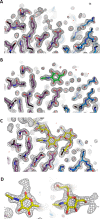
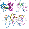
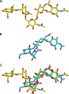
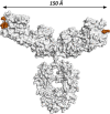
In response to the widespread emergence of antibiotic-resistant microbes, new therapeutic agents are required for many human pathogens. A non-mammalian polysaccharide, poly-N-acetyl-d-glucosamine (PNAG), is produced by bacteria, fungi, and protozoan parasites. Antibodies that bind to PNAG and its deacetylated form (dPNAG) exhibit promising in vitro and in vivo activities against many microbes. A human IgG1 mAb (F598) that binds both PNAG and dPNAG has opsonic and protective activities against multiple microbial pathogens and is undergoing preclinical and clinical assessments as a broad-spectrum antimicrobial therapy. Here, to understand how F598 targets PNAG, we determined crystal structures of the unliganded F598 antigen-binding fragment (Fab) and its complexes with N-acetyl-d-glucosamine (GlcNAc) and a PNAG oligosaccharide. We found that F598 recognizes PNAG through a large groove-shaped binding site that traverses the entire light- and heavy-chain interface and accommodates at least five GlcNAc residues. The Fab-GlcNAc complex revealed a deep binding pocket in which the monosaccharide and a core GlcNAc of the oligosaccharide were almost identically positioned, suggesting an anchored binding mechanism of PNAG by F598. The Fab used in our structural analyses retained binding to PNAG on the surface of an antibiotic-resistant, biofilm-forming strain of Staphylococcus aureus Additionally, a model of intact F598 binding to two pentasaccharide epitopes indicates that the Fab arms can span at least 40 GlcNAc residues on an extended PNAG chain. Our findings unravel the structural basis for F598 binding to PNAG on microbial surfaces and biofilms.
Keywords: Staphylococcus aureus (S. aureus); antibiotic resistance; antibody structure; biofilm; carbohydrate-binding protein; crystal structure; monoclonal antibody; poly-N-acetyl-D-glucosamine; vaccine development.
© 2018 by The American Society for Biochemistry and Molecular Biology, Inc.
Conflict of interest statement
G. B. P. is an inventor of intellectual properties (human monoclonal antibody to PNAG and PNAG vaccines) that are licensed by Brigham and Women's Hospital to Alopexx Vaccine, LLC, and Alopexx Pharmaceuticals, LLC, entities in which G. B. P. also holds equity. As an inventor of intellectual properties, G. B. P. also has the right to receive a share of licensing-related income (royalties, fees) through Brigham and Women's Hospital from Alopexx Pharmaceuticals, LLC, and Alopexx Vaccine, LLC. G. B. P.'s interests were reviewed and are managed by the Brigham and Women's Hospital and Partners Healthcare in accordance with their conflict of interest policies. G. B. P. has received funding from the Morris Animal Foundation and National Institutes of Health to conduct studies on the development of vaccines and antibody therapies targeting the microbial antigen PNAG. C. C.-B. is an inventor of intellectual properties (use of human monoclonal antibody to PNAG and use of PNAG vaccines) that are licensed by Brigham and Women's Hospital to Alopexx Vaccine, LLC, and Alopexx Pharmaceuticals, LLC. As an inventor of intellectual properties, C. C.-B. has the right to receive a share of licensing-related income (royalties, fees) through Brigham and Women's Hospital from Alopexx Pharmaceuticals, LLC, and Alopexx Vaccine, LLC
Figures


References
-
- Roca I., Akova M., Baquero F., Carlet J., Cavaleri M., Coenen S., Cohen J., Findlay D., Gyssens I., Heure O. E., Kahlmeter G., Kruse H., Laxminarayan R., Liébana E., López-Cerero L., et al. (2015) The global threat of antimicrobial resistance: science for intervention. New Microbes New Infect. 6, 22–29 10.1016/j.nmni.2015.02.007 - DOI - PMC - PubMed
-
- Coleman J. P., and Smith C. J. (2011) Microbial resistance, in xPharm: the Comprehensive Pharmacology Reference (Enna S. J., and Bylund D. B., eds) pp. 1–3, Elsevier, Amsterdam
-
- World Health Organization (2017) Global Priority List of Antibiotic-resistant Bacteria to Guide Research, Discovery, and Development of New Antibiotics, World Health Organization, Geneva
Publication types
MeSH terms
Substances
Associated data
- Actions
- Actions
- Actions
- Actions
- Actions
- Actions
- Actions
- Actions
- Actions
LinkOut - more resources
Full Text Sources
Other Literature Sources
Molecular Biology Databases

