Comparative Study
. 2018 Mar;29(3):825-840.
doi: 10.1681/ASN.2017091036. Epub 2018 Feb 15.

Conserved and Divergent Molecular and Anatomic Features of Human and Mouse Nephron Patterning

Affiliations
Comparative Study

Conserved and Divergent Molecular and Anatomic Features of Human and Mouse Nephron Patterning

Nils O Lindström et al. J Am Soc Nephrol. 2018 Mar.

Abstract

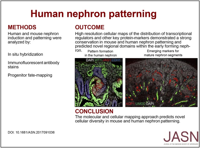
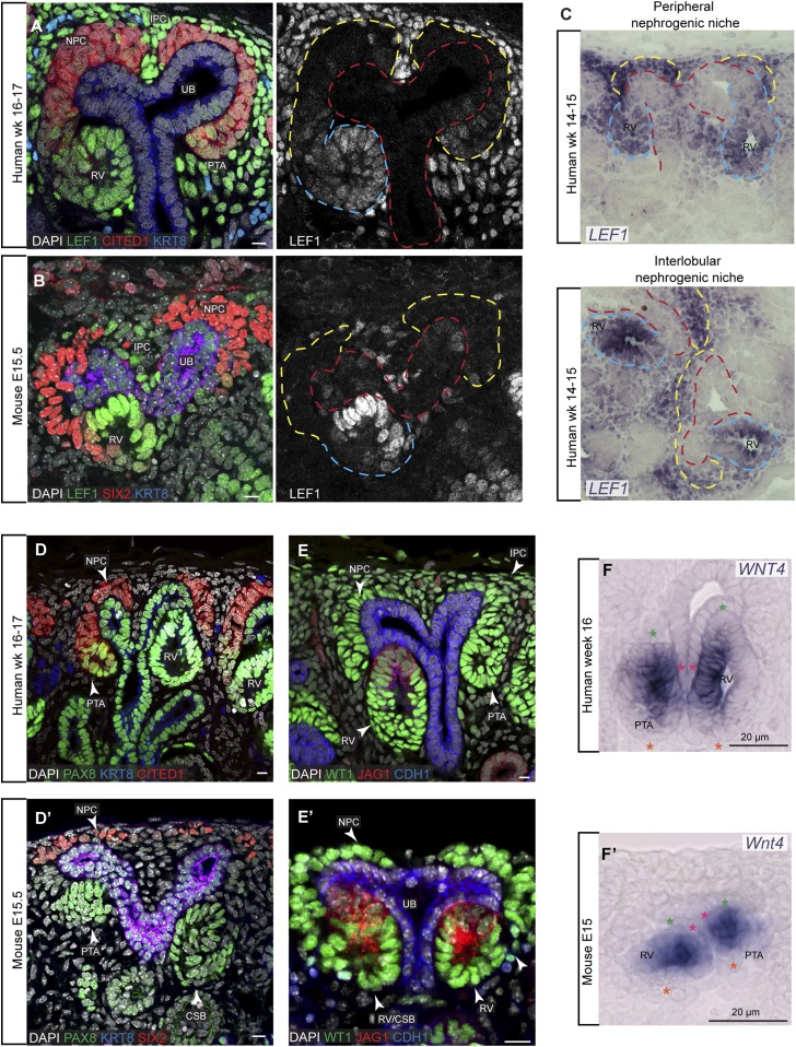
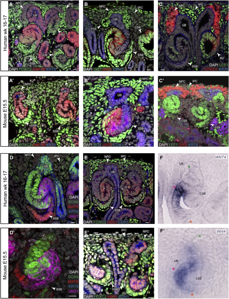
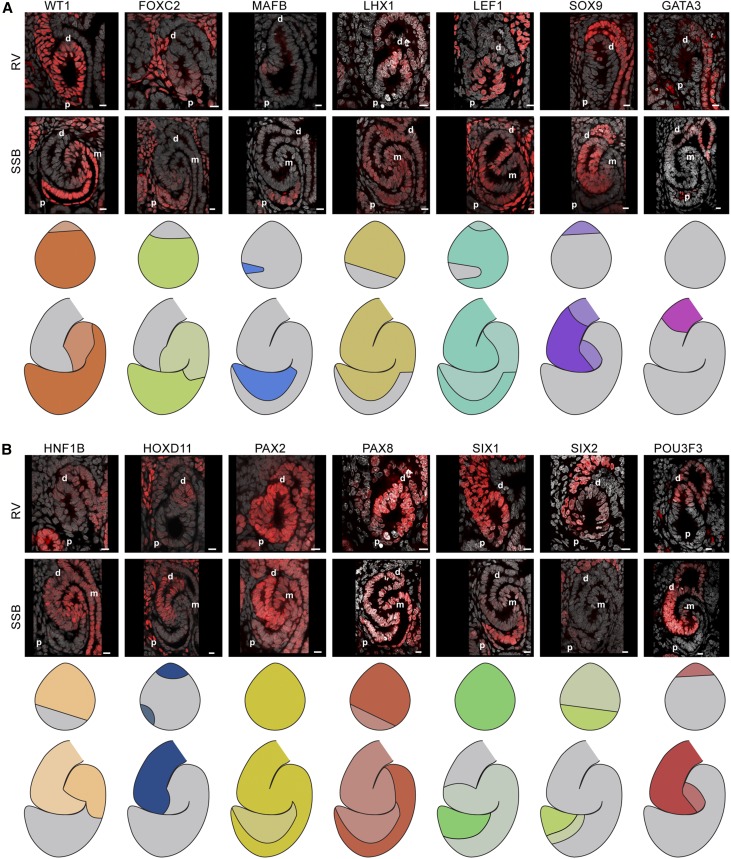
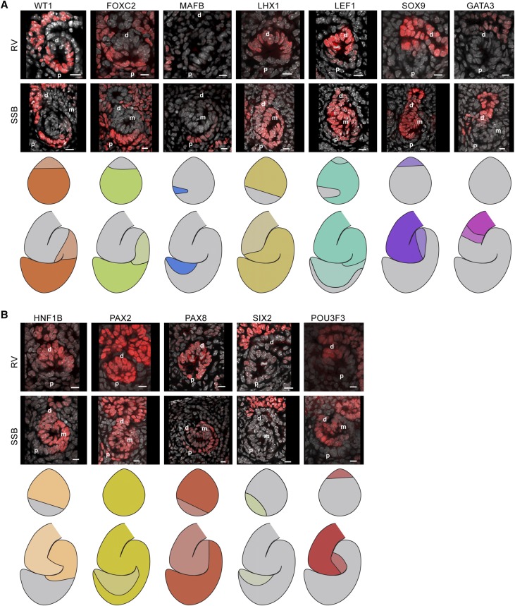
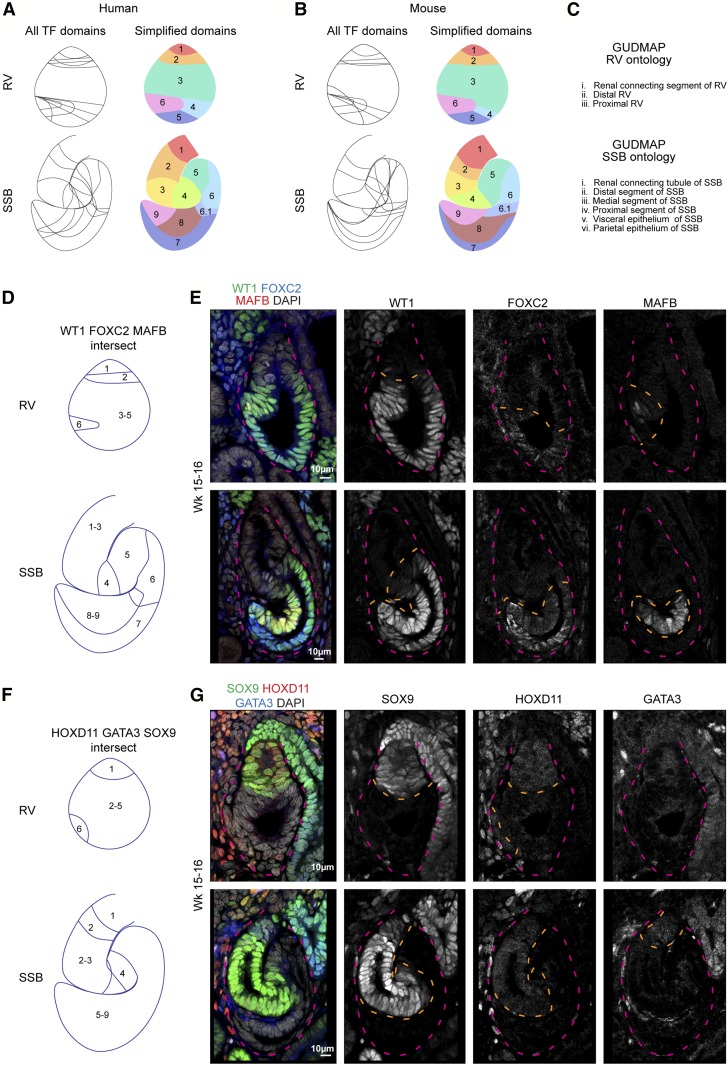
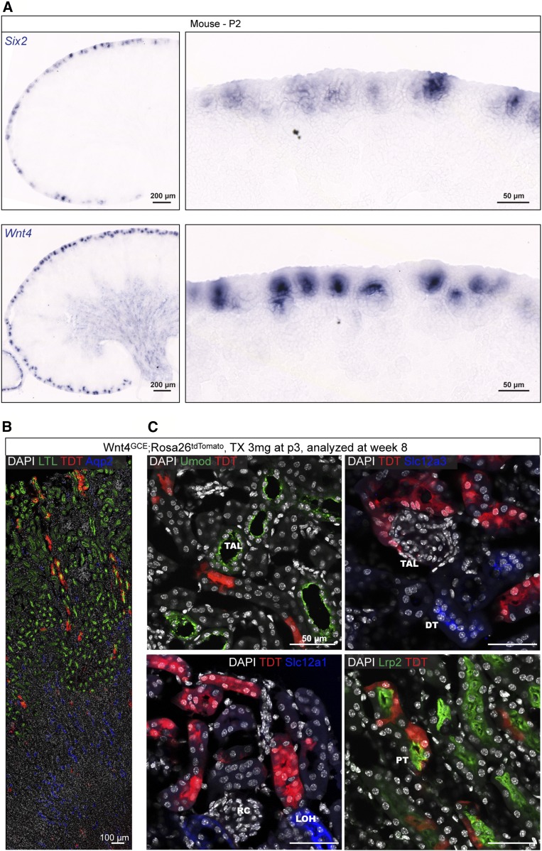
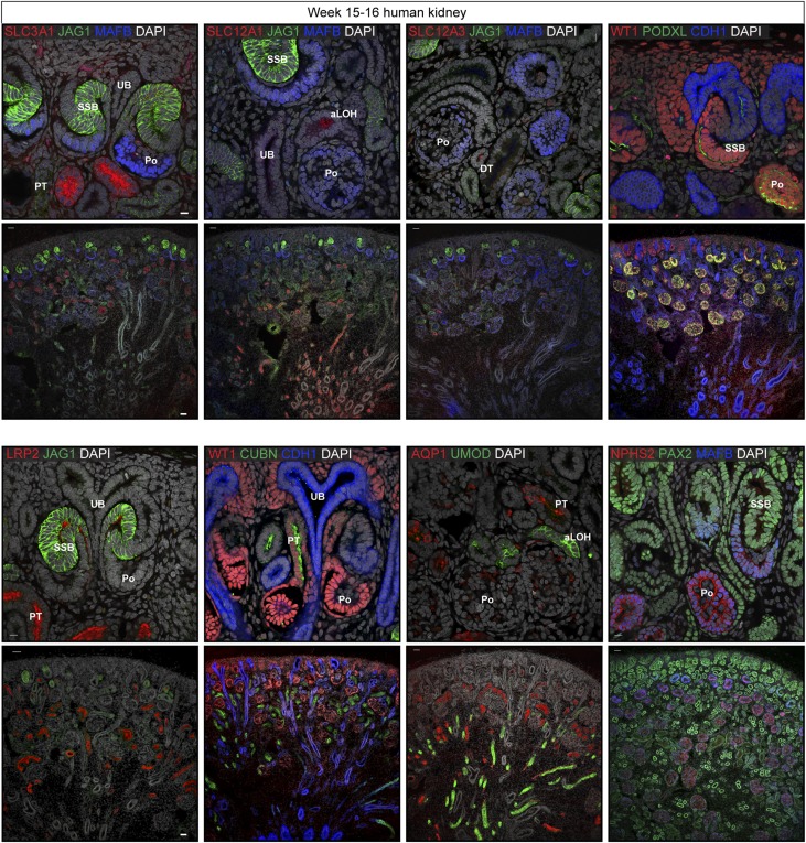
The nephron is the functional unit of the kidney, but the mechanism of nephron formation during human development is unclear. We conducted a detailed analysis of nephron development in humans and mice by immunolabeling, and we compared human and mouse nephron patterning to describe conserved and divergent features. We created protein localization maps that highlight the emerging patterns along the proximal-distal axis of the developing nephron and benchmark expectations for localization of functionally important transcription factors, which revealed unanticipated cellular diversity. Moreover, we identified a novel nephron subdomain marked by Wnt4 expression that we fate-mapped to the proximal mature nephron. Significant conservation was observed between human and mouse patterning. We also determined the time at which markers for mature nephron cell types first emerge-critical data for the renal organoid field. These findings have conceptual implications for the evolutionary processes driving the diversity of mammalian organ systems. Furthermore, these findings provide practical insights beyond those gained with mouse and rat models that will guide in vitro efforts to harness the developmental programs necessary to build human kidney structures.

Keywords: human genetics; kidney development; nephron.


Figures

None
Graphical abstract
Figure 1.
Figure 1.
Nephron progenitor induction in human and mouse nephrogenic niches. (A–F) and (D’–F’) Immunofluorescent stains and in situ hybridization on human and mouse kidneys, respectively. Ages and stains as specified on fields. Yellow, red, and cyan dashed lines indicate cap mesenchyme, ureteric bud, and nephrons, respectively. Stars in (F) and (F’) indicate the nephron axes: green, distal; orange, proximal; magenta start indicates ureteric bud. Scale bars on immunofluorescence data indicate 10 µm. For single-channel views see Supplemental Figures 1 and 3. CSB, comma-shaped body; IPC, interstitial progenitor cells; UB, ureteric bud.
Figure 2.
Figure 2.
Nephron patterning through to the SSB stage in human and mouse kidneys. (A–F) and (A’–F’) Immunofluorescent stains and in situ hybridization on human and mouse kidneys, respectively. Ages and stains as specified on fields. Stars in (F) and (F’) indicate the nephron axes: green, distal; orange, proximal; magenta indicates the ureteric bud. Scale bars indicate 10 µm. CLN, capillary loop stage nephron; CSB, comma-shaped body; IPC, interstitial progenitor cells; UB, ureteric bud.
Figure 3.
Figure 3.
Transcription factor maps in the human renal vesicle and SSB nephron. (A and B) Single-channel immunofluorescent stains with DAPI showing transcription factor localization patterns in renal vesicles and SSB nephrons. Nephron model schematics indicate where the transcription factor is present; a two-level color scheme used to indicate strong and weak detection where applicable. Scale bars indicate 10 µm. Proximal (p), medial (m), and distal (d) segments indicated on fields.
Figure 4.
Figure 4.
Transcription factor maps in the mouse renal vesicle and SSB nephron. (A and B) Single-channel immunofluorescent stains with DAPI showing transcription factor localization patterns in renal vesicles and SSB nephrons. Nephron model schematics indicate where the transcription factor is present; a two-level color scheme used to indicate strong and weak detection where applicable. Scale bars indicate 10 µm. Proximal (p), medial (m), and distal (d) segments indicated on fields.
Figure 5.
Figure 5.
Diversity in the human and mouse renal vesicle and SSB nephron. (A–C) Predicted cellular diversity in the human and mouse RV and SSB and current ontological terms. (D and E) Predicted and tested subdomains identified by detection of WT1, FOXC2, and MAFB in human nephrons. (F and G) Predicted and tested subdomains identified by detection of SOX9, GATA3, and HOXD11 in human nephrons. Dashed magenta line outlines nephrons. Dashed orange line indicates region where the transcription factor is detected.
Figure 6.
Figure 6.
Fate mapping of SSB nephron Wnt4 expression to an adult nephron segment. (A) In situ hybridization on P2 mouse kidneys for Six2 and Wnt4. (B and C) td-Tomato+ cells in a week 8 mouse kidney post fate-mapping from P3. Immunofluorescent stains as stated on fields. DT, distal tubule; LOH, loop of Henle; PT, proximal tubule; RC, renal corpuscle; TAL, thin ascending limb of the loop of Henle.
Figure 7.
Figure 7.
Activation of mature cell-lineage markers in the early development nephron. Immunofluorescent stains for markers of the distal, proximal, loop of Henle, and renal corpuscle domains of the nephron. Stains as specified on fields. Tissue from a week 5–16 human kidney. Scale bar is 10 µm and 50 µm in higher and lower magnification fields, respectively. aLOH, ascending loop of Henle; DT, distal tubule; Po, podocytes; PT, proximal tubule; UB, ureteric bud.

Comment in

  • The Era of Human Developmental Nephrology.
    Nishinakamura R. Nishinakamura R. J Am Soc Nephrol. 2018 Mar;29(3):705-706. doi: 10.1681/ASN.2017121280. Epub 2018 Feb 15. J Am Soc Nephrol. 2018. PMID: 29449450 Free PMC article. No abstract available.

References

    1. Saxen L: Organogenesis of the Kidney, Cambridge, Cambridge University Press, 1987
    1. McMahon AP: Development of the mammalian kidney. Curr Top Dev Biol 117: 31–64, 2016 - PMC - PubMed
    1. Costantini F, Kopan R: Patterning a complex organ: Branching morphogenesis and nephron segmentation in kidney development. Dev Cell 18: 698–712, 2010 - PMC - PubMed
    1. Kobayashi A, Valerius MT, Mugford JW, Carroll TJ, Self M, Oliver G, McMahon AP: Six2 defines and regulates a multipotent self-renewing nephron progenitor population throughout mammalian kidney development. Cell Stem Cell 3: 169–181, 2008 - PMC - PubMed
    1. Boyle S, Misfeldt A, Chandler KJ, Deal KK, Southard-Smith EM, Mortlock DP, Baldwin HS, de Caestecker M: Fate mapping using Cited1-CreERT2 mice demonstrates that the cap mesenchyme contains self-renewing progenitor cells and gives rise exclusively to nephronic epithelia. Dev Biol 313: 234–245, 2008 - PMC - PubMed

Publication types

MeSH terms