Comparative Study
. 2018 Feb 15;9(3):256.
doi: 10.1038/s41419-018-0294-6.

ALS-related human cortical and motor neurons survival is differentially affected by Sema3A

Affiliations
Comparative Study

ALS-related human cortical and motor neurons survival is differentially affected by Sema3A

Anastasya Birger et al. Cell Death Dis. .

Abstract

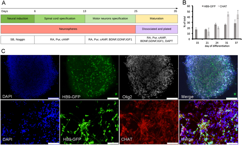
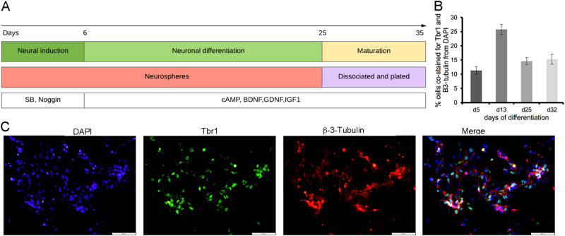
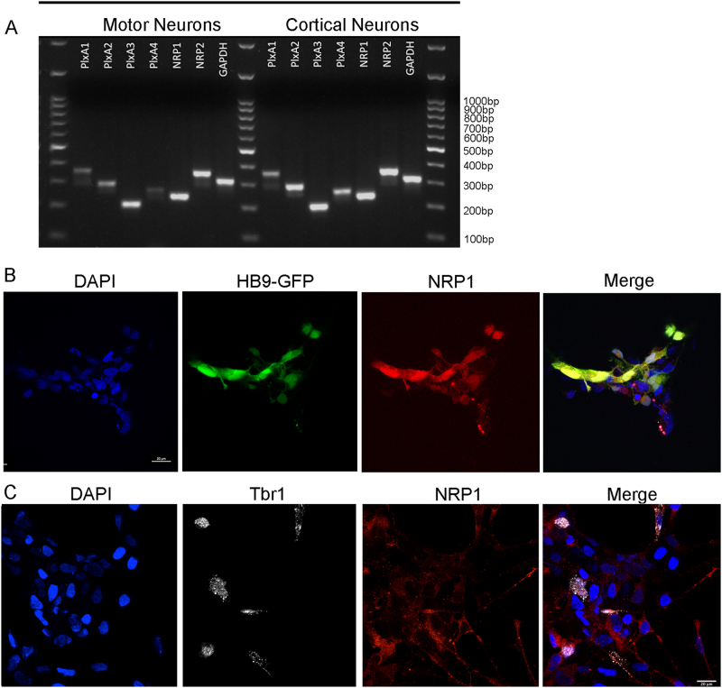
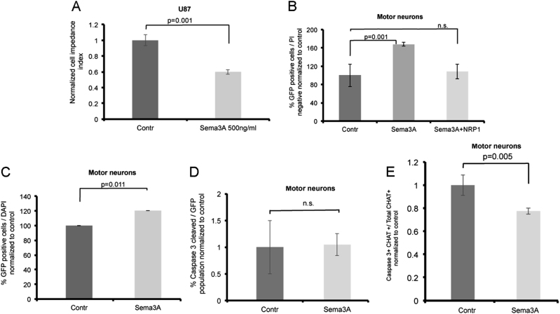
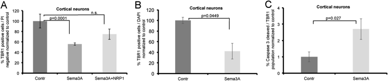
Amyotrophic lateral sclerosis (ALS) is a neurodegenerative disease characterized by cell death of upper and lower motor neurons (MNs). The cause of MN cell loss is not completely understood but involves both cell autonomous and non-cell autonomous mechanisms. Numerous molecules have been implicated to be involved in the death of MNs. One such candidate is semaphorin 3A (Sema3A). In ALS patients, Sema3A was shown to be significantly upregulated in the motor cortex and downregulated in the spinal cord. In the mouse, Sema3A was shown to be an axon repellent molecule for MNs. Sema3A could also induce death of different neuronal types that are also repelled by it, including sensory, sympathetic, retinal, and cortical neurons. In contrast, astrocyte-specific knockout of Sema3A results in motor neuron cell death, consistent with the idea that Sema3A is a survival factor for mouse motor neurons. Here, we tested the response of human cortical neurons and spinal cord MNs to Sema3A. We found that Sema3A enhances the survival of spinal cord MNs. In contrast, Sema3A reduces the survival of cortical neurons. Thus, both upregulation of Sema3A in the cortex, or downregulation in the spinal cord of ALS patients is likely to directly contribute to MNs cell loss in ALS patients.


Conflict of interest statement

The authors declare that they have no conflict of interest.

Figures

Fig. 1
Fig. 1. hESC-derived spinal motor neuron.
a Graphical representation of hESC differentiation into MNs. b, c hESC monitored at different time points of differentiation. b Quantification of cells positive for GFP expressed under the promoter of early MNs marker HB9 and immunostained for the mature MN marker CHAT at different time points of the protocol. Data are represented as mean ± SEM of three independent experiments. c Representative fluorescence microscopy images at days 15 (upper line, scale bars = 200 µm) and day 31 (lower line, scale bars = 100 µm) are shown.
Fig. 2
Fig. 2. hESC-derived cortical neurons.
a Graphical representation of hESC differentiation into cortical neurons. b Quantification of cells positive for Tbr1 at different time points of the protocol. Data are represented as mean ± SEM of three independent experiments. c Neurons at days 30 of the differentiation protocol were immunostained for Trb1 (cortical marker) and β-3-tubulin (a general neuronal marker). Representative confocal microscopy images are shown (scale bars = 100 µm).
Fig. 3
Fig. 3. MNs and cortical neurons express the Sema3A receptors.
a RT-PCR analysis for 1-PlexinA1, 2-PlxinA2, 3-PlxinA3, 4-PlxinA4, 5-NRP1, 6-NRP2, 7-GAPDH, and M-DNA ladder 100 bp. b, c MNs (b) and cortical neurons (c) were immunostained and imaged at days 30. Representative confocal microscopy images are shown (scale bars = 20 µm (b) and bars = 25 µm (c).
Fig. 4
Fig. 4. Sema3A improves the survival of human spinal motor neurons.
a Cell-electrode impedance assay indicates response of U87MG cells to Sema3A. b Percentage of HB9-GFP MNs was analyzed by FACS 72 h after treatment with Sema3A, Sema3A with NRP1 blocking antibody or control media. MNs were gated as PI-negative and GFP-positive relative to GFP-negative control cells. Data are normalized to control treatment and represented as mean of three independent experiment ± SEM. P value calculated by unpaired t test. c Quantification of imaged HB9-GFP-positive MNs 48 h after treatment with Sema3A or control media. Data are represented as mean ± SEM of four independent experiments. P value calculated by unpaired t test. d Quantification of immunostained and imaged MNs positive for GFP and cleaved caspase-3. Data are represented as mean ± SEM of three independent experiments. e Quantification of immunostained and imaged MNs positive for CHAT and cleaved caspase-3. Data are represented as mean ± SEM of three independent experiments. P value calculated by unpaired t test.
Fig. 5
Fig. 5. Sema3A induces the death of human cortical neurons.
a The percentage of cortical neurons was analyzed by FACS 72 h after treatment with Sema3A, Sema3A with NRP1 blocking antibody or control media. Cortical neurons were immunostained and gated as PI-negative and Tbr1-positive relative to unstained control cells. Data are normalized to control treatment and represented as mean of three independent experiment ± SEM. P value calculated by unpaired t test. b Quantification of imaged cortical neurons stained for Tbr1 after 72 h treatment with Sema3A and control media. c Quantification of immunostained and imaged cortical neurons positive for Tbr1 and cleaved caspase-3. Data are represented as mean ± SEM of three independent experiments.

References

    1. Peters OM, Ghasemi M, Brown RH. Emerging mechanisms of molecular pathology in ALS. J. Clin. Invest. 2015;125:1767–1779. doi: 10.1172/JCI71601. - DOI - PMC - PubMed
    1. Taylor JP, Brown RH, Jr, Cleveland DW. Decoding ALS: from genes to mechanism. Nature. 2016;539:197–206. doi: 10.1038/nature20413. - DOI - PMC - PubMed
    1. Huber AB, et al. Distinct roles for secreted semaphorin signaling in spinal motor axon guidance. Neuron. 2005;48:949–964. doi: 10.1016/j.neuron.2005.12.003. - DOI - PubMed
    1. Shirvan A, et al. Anti-semaphorin 3A antibodies rescue retinal ganglion cells from cell death following optic nerve axotomy. J. Biol. Chem. 2002;277:49799–49807. doi: 10.1074/jbc.M204793200. - DOI - PubMed
    1. Ben-Zvi A, et al. The semaphorin receptor plexinA3 mediates neuronal apoptosis during dorsal root ganglia development. J. Neurosci. 2008;28:12427–12432. doi: 10.1523/JNEUROSCI.3573-08.2008. - DOI - PMC - PubMed

Publication types