. 2018 Feb 15;9(3):267.
doi: 10.1038/s41419-018-0332-4.

Glucocorticoids promote apoptosis of proinflammatory monocytes by inhibiting ERK activity

Affiliations

Glucocorticoids promote apoptosis of proinflammatory monocytes by inhibiting ERK activity

Adrian Achuthan et al. Cell Death Dis. .

Abstract

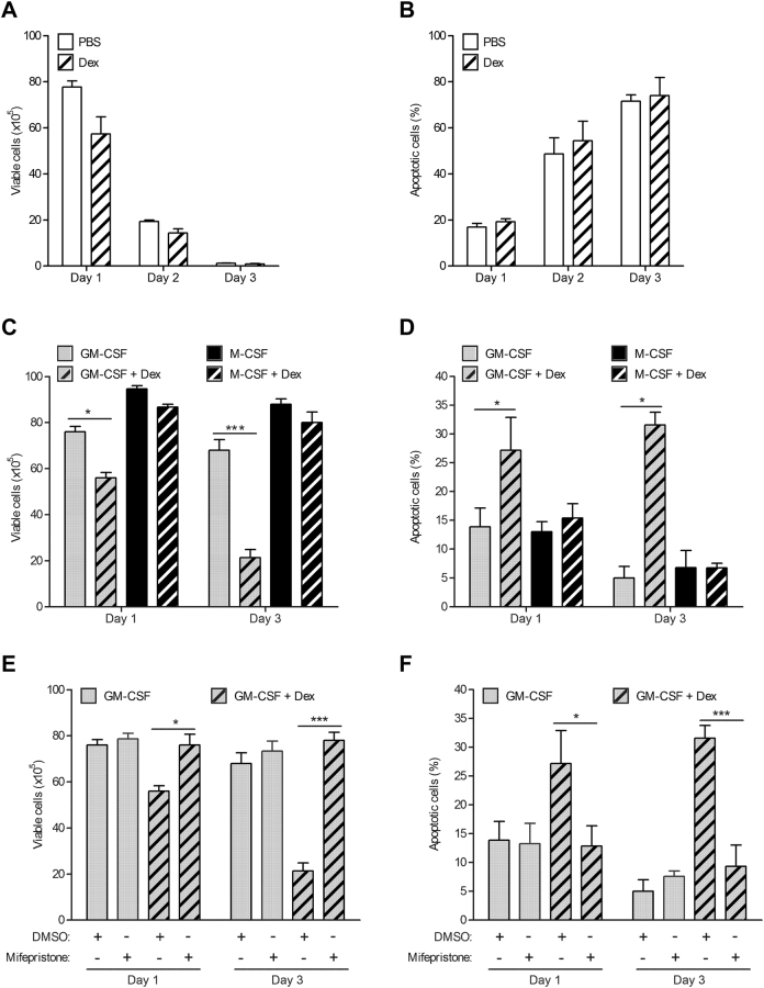
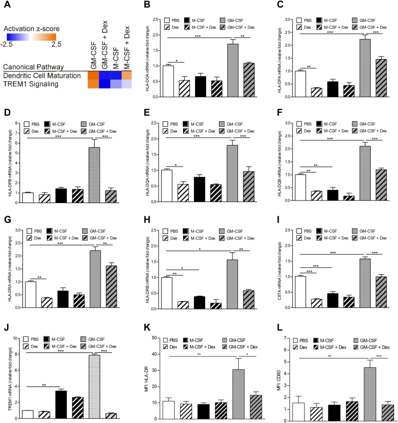
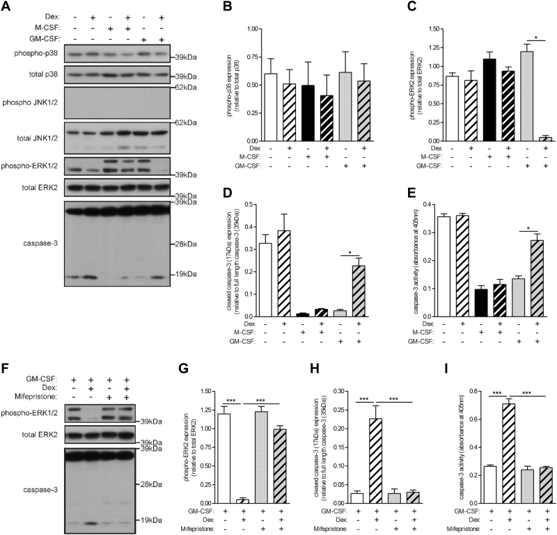
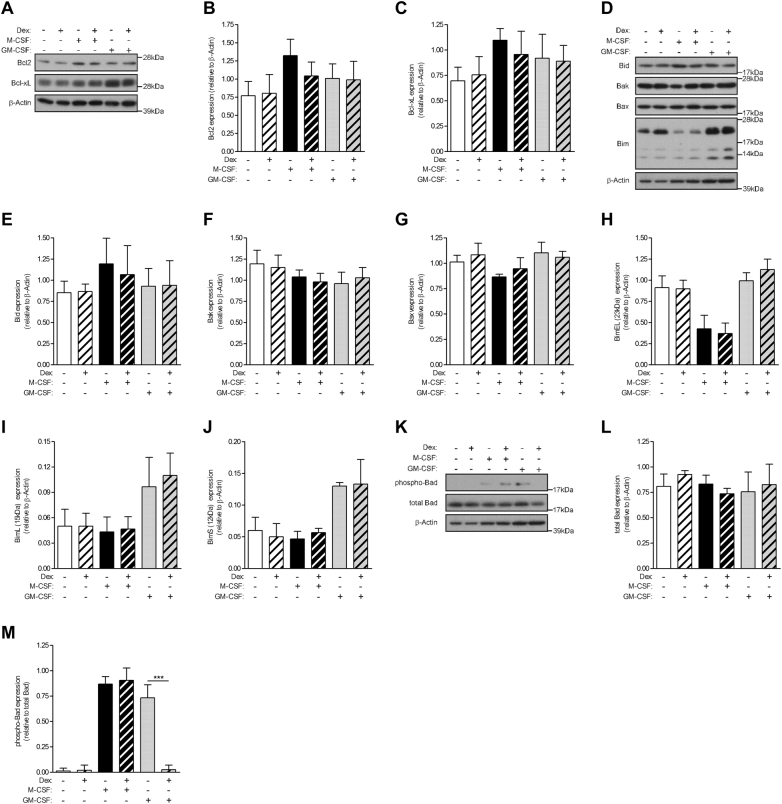
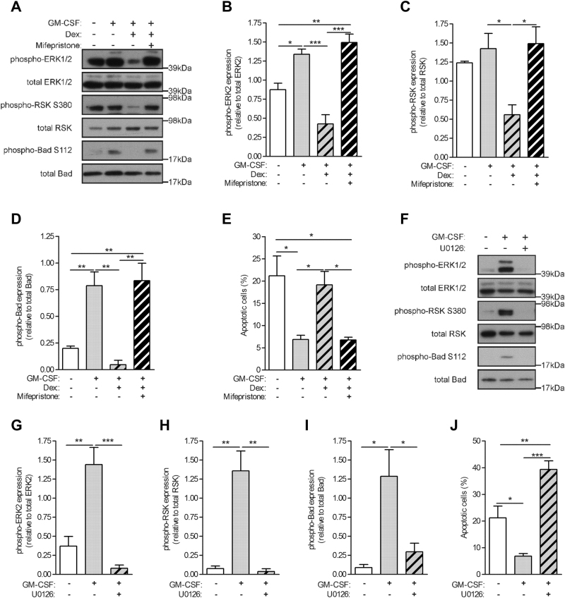
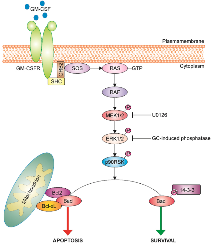
Glucocorticoids (GCs) are potent anti-inflammatory drugs whose mode of action is complex and still debatable. One likely cellular target of GCs are monocytes/macrophages. The role of GCs in monocyte survival is also debated. Although both granulocyte macrophage-colony stimulating factor (GM-CSF) and macrophage-CSF (M-CSF) are important regulators of macrophage lineage functions including their survival, the former is often associated with proinflammatory functions while the latter is important in lineage homeostasis. We report here that the GC, dexamethasone, induces apoptosis in GM-CSF-treated human monocytes while having no impact on M-CSF-induced monocyte survival. To understand how GCs, GM-CSF, and M-CSF are regulating monocyte survival and other functions during inflammation, we firstly examined the transcriptomic changes elicited by these three agents in human monocytes, either acting alone or in combination. Transcriptomic and Ingenuity pathway analyses found that dexamethasone differentially modulated dendritic cell maturation and TREM1 signaling pathways in GM-CSF-treated and M-CSF-treated monocytes, two pathways known to be regulated by ERK1/2 activity. These analyses led us to provide evidence that the GC inhibits ERK1/2 activity selectively in GM-CSF-treated monocytes to induce apoptosis. It is proposed that this inhibition of ERK1/2 activity leads to inactivation of p90 ribosomal-S6 kinase and Bad dephosphorylation leading in turn to enhanced caspase-3 activity and subsequent apoptosis. Furthermore, pharmacological inhibition of GC receptor activity restored the ERK1/2 signaling and prevented the GC-induced apoptosis in GM-CSF-treated monocytes. Increased tissue macrophage numbers, possibly from enhanced survival due to mediators such as GM-CSF, can correlate with inflammatory disease severity; also reduction in these numbers can correlate with the therapeutic benefit of a number of agents, including GCs. We propose that the ERK1/2 signaling pathway promotes survival of GM-CSF-treated proinflammatory monocytes, which can be selectively targeted by GCs as a novel mechanism to reduce local monocyte/macrophage numbers and hence inflammation.


Conflict of interest statement

The authors declare that they have no conflict of interest.

Figures

Fig. 1
Fig. 1. Glucocorticoid induces apoptosis in GM-CSF-treated monocytes but not in M-CSF-treated monocytes.
a, b Human monocytes (1 × 106) were cultured in the absence (PBS) or presence of dexamethasone (Dex) (100 nM) over three days. a Viable cells b apoptotic cells by trypan blue exclusion method and Annexin V staining, respectively. c, d Human monocytes (1 × 106) were cultured with either GM-CSF (10 ng/ml) alone, M-CSF (5,000 U/ml) alone or together with Dex over three day period. c Viable cells d apoptotic cells were determined. ad Experiments with the same donors. e, f Human monocytes (1 × 106) were pre-treated with mifepristone (1 μM) for 30 min before culture in either GM-CSF alone or together with Dex over three day period. e Viable cells f apoptotic cells were determined. Graphs were plotted as mean ± SEM (N = 4). Statistical analyses were performed using Student’s paired t-test, where *p < 0.05 and ***p < 0.001
Fig. 2
Fig. 2. Glucocorticoid inhibits GM-CSF-regulated dendritic cell maturation and TREM1 signaling pathways.
Human monocytes (1 × 106) were treated with either dexamethasone (Dex) (100 nM), M-CSF (5000 U/ml), GM-CSF (10 ng/ml), or as a combination of Dex with each CSF for 16 h. Isolated RNA was subjected to a gene expression arrays, showing significantly regulated canonical pathways among the treatment groups by ingenuity pathway comparison analysis. bj qPCR with HPRT RNA as reference. mRNA expression, assayed in triplicate, was plotted relative to that at 16 h in culture medium containing vehicle (PBS), which was given an arbitrary value of 1.0. FACS analysis of cell surface expression of k HLA-DR and l CD80. The graphs represent mean ± SEM (N = 4). Statistical analyses were performed using one-way ANOVA with Tukey’s multiple comparison test, where *p < 0.05, **p < 0.01, and ***p < 0.001
Fig. 3
Fig. 3. Glucocorticoid inhibits GM-CSF-activated ERK1/2 phosphorylation, which is accompanied by increased caspase-3 activity.
ae Human monocytes (1 × 106) were treated with either dexamethasone (Dex) (100 nM), M-CSF (5000 U/ml), GM-CSF (10 ng/ml), or as a combination of Dex with each CSF for 16 h. a Whole-cell lysates were subjected to Western blotting with anti-phospho-p38, anti-p38, anti-phospho-JNK, anti-JNK, anti-phospho-ERK1/2, anti-ERK2, and anti-caspase-3 Abs. bd Quantified data of activated/cleaved protein relative to total/full-length protein, as indicated e Caspase-3 activity. fi Human monocytes (1 × 106) were pre-treated with mifepristone (1 μM) for 30 min before treated with either GM-CSF alone or together with Dex for 16 h. f Whole-cells lysates were subjected to Western blotting with anti-phospho-ERK1/2, anti-ERK2 and anti-caspase-3 Abs. g, h Quantified data of activated/cleaved protein relative to total/full-length protein, as indicated i Caspase-3 activity. ai Experiments from the same donors. Graphs were plotted as mean ± SEM (N = 3). Statistical analyses were performed using one-way ANOVA with Tukey’s multiple comparison test; *p < 0.05 and ***p < 0.001
Fig. 4
Fig. 4. Glucocorticoid inhibits GM-CSF-induced Bad phosphorylation.
Human monocytes (1 × 106) were treated with either dexamethasone (Dex) (100 nM), M-CSF (5000 U/ml), GM-CSF (10 ng/ml), or as a combination of Dex with each CSF for 16 h. Whole-cell lysates were subjected to Western blotting with antibodies against a anti-apoptotic proteins d pro-apoptotic proteins k Bad phosphorylation. b, c, ej, l, m Quantified data of activated/apoptotic protein relative to total/β-actin protein, as indicated. Graphs were plotted as mean ± SEM (N = 3). Statistical analyses were performed using one-way ANOVA with Tukey’s multiple comparison test; ***p < 0.001
Fig. 5
Fig. 5. Glucocorticoid-mediated inhibition of ERK1/2 activity leads to Bad dephosphorylation and monocyte apoptosis.
ae Human monocytes (1 × 106) were pre-treated with mifepristone (1 μM) or DMSO for 30 min before treatment with GM-CSF (10 ng/ml) alone or together with dexamethasone (Dex) (100 nM) for 16 h. a Whole-cell lysates were subjected to Western blotting with anti-phospho-ERK1/2, total ERK1/2, phospho-RSK, total RSK, phospho-Bad, or total Bad Abs. bd Quantified data of activated protein relative to total protein, as indicated. e Apoptotic cells by Annexin V staining. fj Human monocytes (1 × 106) were pre-treated with MEK inhibitor, UO126 (10 μM), or DMSO for 30 min before treatment with GM-CSF (10 ng/ml) for 16 h. f Whole-cell lysates were subjected to Western blotting with anti-phospho-ERK1/2, total ERK1/2, phospho-RSK, total RSK, phospho-Bad, or total Bad Abs. gi Quantified data of activated protein relative to total protein, as indicated. j Apoptotic cells by Annexin V staining. Graphs were plotted as mean ± SEM (N = 3). Statistical analyses were performed using one-way ANOVA with Tukey post-test; *p < 0.05, **p < 0.01, and ***p < 0.001
Fig. 6
Fig. 6. Proposed glucocorticoid-induced apoptotic signaling pathway in GM-CSF-treated monocytes.
A schematic diagram illustrating a possible GM-CSF-induced ERK1/2 signaling pathway in monocytes which is inhibited by a GC leading to Bad deactivation and subsequent apoptosis. GM-CSF binding to its receptor activates a Raf/MEK/ERK signaling cascade leading in turn to ribosomal associated p90RSK kinase activation. The pro-apoptotic Bad protein is phosphorylated by p90RSK and subsequently binds to the cytoskeletal scaffold protein, 14-3-3. In the absence of a growth factor or upon inhibition of the Raf/MEK/ERK pathway by the pharmacological inhibitor (U0126) or a GC-induced phosphatase(s), p90RSK remains inactivated and is unable to phosphorylate Bad. Non-phosphorylated Bad in turn can interfere with the anti-apoptotic proteins, Bcl-xL and Bcl2, resulting in loss of mitochondrial membrane integrity and ultimately apoptosis

References

    1. Straub RH, Cutolo M. Glucocorticoids and chronic inflammation. Rheumatology. 2016;55:ii6–ii14. doi: 10.1093/rheumatology/kew348. - DOI - PubMed
    1. Barnes PJ. Corticosteroids: the drugs to beat. Eur. J. Pharmacol. 2006;533:2–14. doi: 10.1016/j.ejphar.2005.12.052. - DOI - PubMed
    1. Newton R. Anti-inflammatory glucocorticoids: Changing concepts. Eur. J. Pharmacol. 2013;724:231–236. doi: 10.1016/j.ejphar.2013.05.035. - DOI - PubMed
    1. Rhen T, Cidlowski JA. Anti-inflammatory action of glucocorticoids—new mechanisms for old drugs. N. Engl. J. Med. 2005;353:1711–1723. doi: 10.1056/NEJMra050541. - DOI - PubMed
    1. Burgess AW, Metcalf D. The nature and action of granulocyte-macrophage colony stimulating factors. Blood. 1980;56:947–958. - PubMed

Publication types

MeSH terms

Substances