. 2018 Feb 15;8(1):3118.
doi: 10.1038/s41598-018-21384-z.

Intravenous synthetic platelet (SynthoPlate) nanoconstructs reduce bleeding and improve 'golden hour' survival in a porcine model of traumatic arterial hemorrhage

Affiliations

Intravenous synthetic platelet (SynthoPlate) nanoconstructs reduce bleeding and improve 'golden hour' survival in a porcine model of traumatic arterial hemorrhage

DaShawn A Hickman et al. Sci Rep. .

Abstract

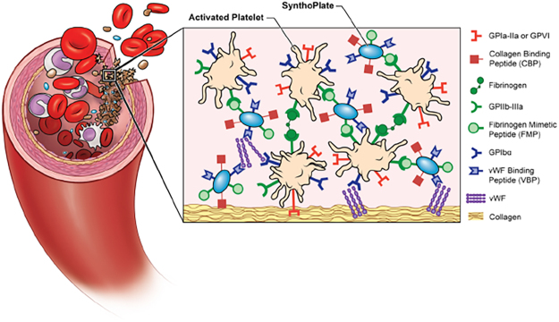
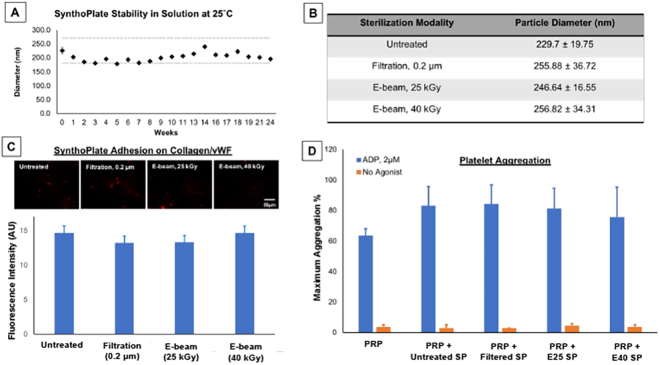
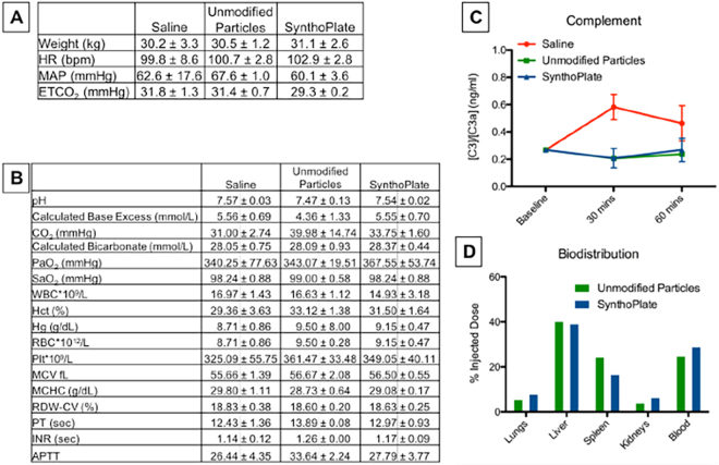
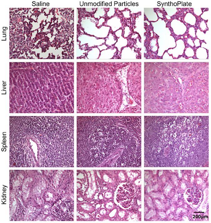
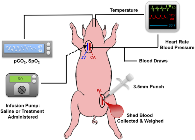
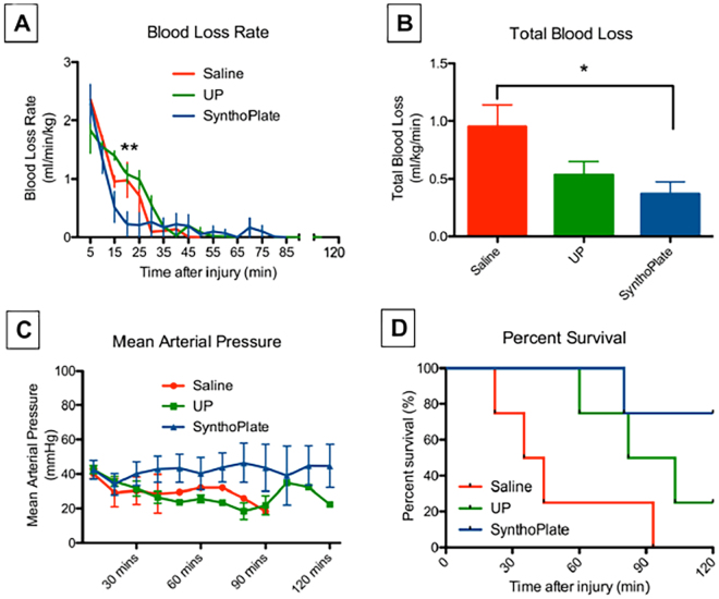
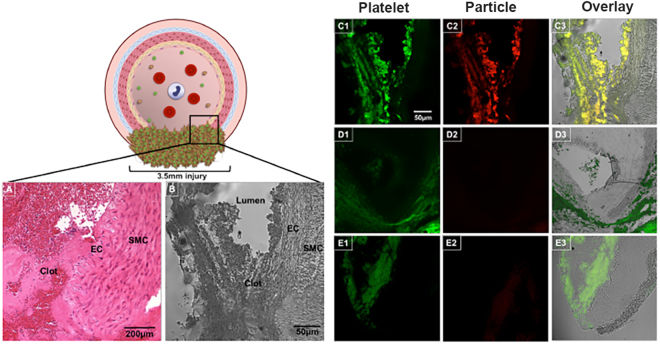
Traumatic non-compressible hemorrhage is a leading cause of civilian and military mortality and its treatment requires massive transfusion of blood components, especially platelets. However, in austere civilian and battlefield locations, access to platelets is highly challenging due to limited supply and portability, high risk of bacterial contamination and short shelf-life. To resolve this, we have developed an I.V.-administrable 'synthetic platelet' nanoconstruct (SynthoPlate), that can mimic and amplify body's natural hemostatic mechanisms specifically at the bleeding site while maintaining systemic safety. Previously we have reported the detailed biochemical and hemostatic characterization of SynthoPlate in a non-trauma tail-bleeding model in mice. Building on this, here we sought to evaluate the hemostatic ability of SynthoPlate in emergency administration within the 'golden hour' following traumatic hemorrhagic injury in the femoral artery, in a pig model. We first characterized the storage stability and post-sterilization biofunctionality of SynthoPlate in vitro. The nanoconstructs were then I.V.-administered to pigs and their systemic safety and biodistribution were characterized. Subsequently we demonstrated that, following femoral artery injury, bolus administration of SynthoPlate could reduce blood loss, stabilize blood pressure and significantly improve survival. Our results indicate substantial promise of SynthoPlate as a viable platelet surrogate for emergency management of traumatic bleeding.


Conflict of interest statement

A. Sen Gupta is a co-inventor on patents related to SynthoPlate technology: US 9107845 and US 9,6363,383. The SynthoPlate trademark is currently recorded with USPTO (U.S. Serial Number: 86–829,160). The other authors state that they have no conflict of interest.

Figures

Figure 1
Figure 1
Schematic representation of SynthoPlate design and mechanism, showing nanoconstructs heteromutlivalently decorated with VBP, CBP and FMP motifs to render platelet-mimetic interactions with vWF, collagen and active platelet integrin GPIIb-IIIa respectively, and thus amplify platelet-mediated primary hemostatic mechanisms at the injury site.
Figure 2
Figure 2
[A] SynthoPlate diameter (size distribution) analysis over a 6-month period demonstrated that the particles retained their size within 20% of their starting diameter when stored in a 0.9% NaCl solution at 25 °C, indicating long term stability; [B] After sterilization with filtration or E-beam, SynthoPlate showed minimal alteration in size (diameter) compared to fresh-made (unsterilized) samples, indicating that the sterilization did not affect particle stability; [C] Representative fluorescent images and quantitative analysis of surface-averaged fluorescence intensity of adhered SynthoPlate showing that sterilized SynthoPlate (red) maintained its ability to adhere to ‘collagen + vWF’-coated surface at levels similar to fresh-made (unsterilized) SynthoPlate, indicating that sterilization did not affect the biofunctional properties of VBP and CBP; [D] Aggregometry analysis showed that neither fresh-made (unsterilized) or sterilized SynthoPlate induced spontaneous aggregation of platelets without agonist (w/o ADP), but both unsterilized and sterilized SynthoPlate markedly increased platelet aggregation in the presence of ADP, indicating that sterilization did not affect the biofunctional property of FMP on SynthoPlate and also SynthoPlate did not have any thrombotic risk towards resting platelets.
Figure 3
Figure 3
[A] Average vitals over the course of the experiment of uninjured pigs and [B] average blood lab values over the course of the experiment of uninjured pigs upon administration of unmodified particles or SynthoPlate, showed that there are no statistically significant differences in values compared to saline administration, indicating that SynthoPlate administration has no detrimental physiological effect; [C] Analysis of C3:C3a plasma concentration ratios in blood drawn from pigs administered with unmodified particles or SynthoPlate showed no statistically significant alterations of this ratio compared to saline administration groups, indicating that in the dose and administration protocol used, SynthoPlate did not have complement activation (and CARPA) risk; [D] Biodistribution analysis from harvested organs indicated a similar distribution profile for unmodified particles and SynthoPlate, with a majority of particles cleared through the liver.
Figure 4
Figure 4
Representative hemotoxylin and eosin (H&E) stained histology images (32× magnification) of organ samples from pigs treated with saline, unmodified particles or SynthoPlate particles, show that SynthoPlate (as well as unmodified particles) does not cause any thrombotic risks in organs; scale bar of 200 μm shown in bottom row kidney image is applicable to all histology images.
Figure 5
Figure 5
Schematic representation of pig femoral artery hemorrhage model setup. A CO2 sensor was placed at the end of the endotracheal tube and mechanical ventilation was provided, EKG electrodes were placed on the pig’s limbs, a pulse-oximeter probe was placed on the pig’s mouth, an esophageal temperature probe was placed to measure core temperature, an angiocatheter was placed in the carotid artery to acquire invasive blood pressure and also withdraw blood samples for ex vivo analysis, an angiocatheter was placed in the internal jugular vein to deliver saline (or nanoparticle treatments) via an infusion pump.
Figure 6
Figure 6
Hemostatic efficacy analysis in injured pigs shows that [A] pigs administered with SynthoPlate had a reduced blood loss rate (blue) compared to those treated with saline (red) and unmodified particles (UP, green), especially within first 30 min post treatment administration, where blood loss rate in SynthoPlate treated pig was significantly lower than those treated with unmodified (control) particles and saline (**p < 0.01); [B] SynthoPlate administration in pigs also resulted in significantly lower total blood loss compared to saline administration (*p < 0.05); [C] SynthoPlate administration in pigs resulted in maintenance and stabilization of a higher average mean arterial blood pressure (MAP) over time (data points shown for every 10 min); [D] SynthoPlate administration in pigs resulted in a significant enhancement of survival, with 100% surviving the first 60 min (golden hour) and 75% surviving the additional 60 min (p < 0.05), compared to administration of saline (25% survival by 60 min and 0% survival by ~90 min) or unmodified particles (50% survival by 90 min, 25% survival at 120 min).
Figure 7
Figure 7
Schematic representation and representative images of hemostasized injury site in the femoral artery of pigs treated with SynthoPlate or saline or control particles: [A] Representative hematoxylin and eosin (H&E) stained histology image (32× magnification) and [B] representative bright field image (10× magnification) of the site of injury (transected artery) with the injured vessel components (EC: endothelial cell, SMC: smooth muscle cell) and hemostatic clot in view for injured pig treated with SynthoPlate; [C1] representative fluorescence image (10× magnification) of FITC-anti-CD42b (green fluorescence) stained platelets, [C2] Rhodamine B-labeled (red fluorescence) SynthoPlate particles and [C3] overlay of C1 and C2, in the same field of view as the brightfield image B demonstrating that red fluorescent SynthoPlate is co-localized with green fluorescent platelets at the site of injury within the hemostatic clot; [D1, D2 and D3] are similar representative images for injured pig treated with saline, and [E1, E2 and E3] are similar representative images for injured pig treated with unmodified (control) particles showing that some platelets indeed localize at the injury site to promote hemostasis but presence of control particle is minimal at the site and therefore control particles do not have the ability to augment hemostasis; scale bar of 50 μm shown in C1 is applicable to all fluorescent images.

References

    1. Kauvar DS, Lefering R, Wade CE. Impact of Hemorrhage on Trauma Outcome: An Overview of Epidemiology, Clinical Presentations, and Therapeutic Considerations. J Trauma. 2006;60:S3–11. doi: 10.1097/01.ta.0000199961.02677.19. - DOI - PubMed
    1. Holcomb JB, et al. Causes of Death in U.S. Special Operations Forces in the Global War on Terrorism, 2001–2004. Ann. Surg. 2007;245:986–991. doi: 10.1097/01.sla.0000259433.03754.98. - DOI - PMC - PubMed
    1. Blackbourne LH, et al. Military medical revolution: prehospital combat casualty care. J. Trauma Acute Care Surg. 2012;76:S372–S377. doi: 10.1097/TA.0b013e3182755662. - DOI - PubMed
    1. Cohen, M.J. et al. PROMMTT Study Group: Clinical and mechanistic drivers of acute traumatic coagulopathy. J. Trauma Acute Care Surg. 75, S40–S47 (2013). - PMC - PubMed
    1. Dorlac WC, et al. Mortality from isolated civilian penetrating injury. J. Trauma. 2005;59:217–222. doi: 10.1097/01.TA.0000173699.71652.BA. - DOI - PubMed

Publication types