. 2018 Feb 1:10:20.
doi: 10.3389/fnagi.2018.00020. eCollection 2018.

Atrial Natriuretic Peptide Acts as a Neuroprotective Agent in in Vitro Models of Parkinson's Disease via Up-regulation of the Wnt/β-Catenin Pathway

Affiliations

Atrial Natriuretic Peptide Acts as a Neuroprotective Agent in in Vitro Models of Parkinson's Disease via Up-regulation of the Wnt/β-Catenin Pathway

Arianna Colini Baldeschi et al. Front Aging Neurosci. .

Abstract

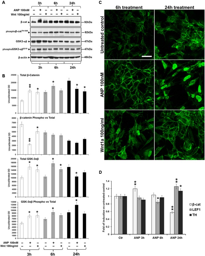
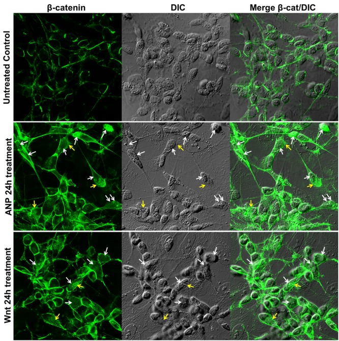
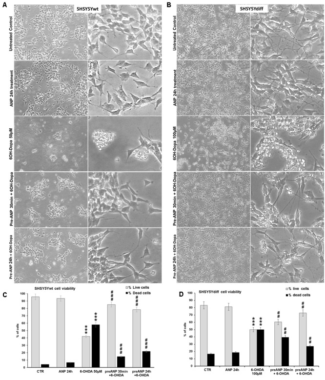
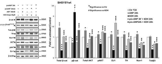
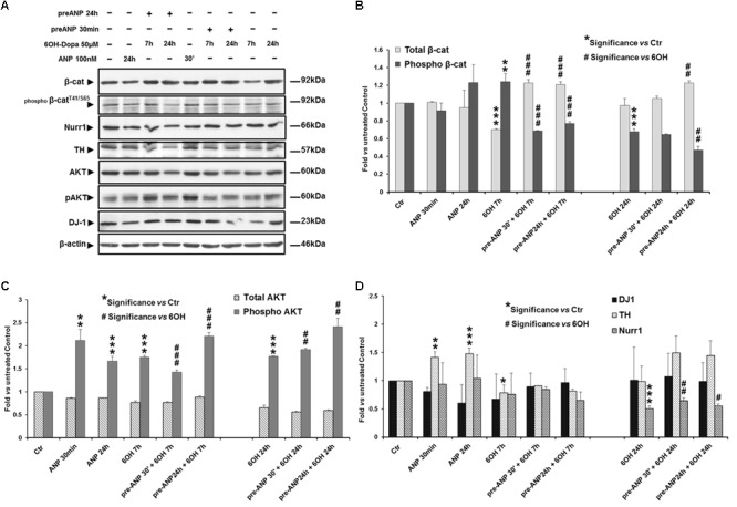
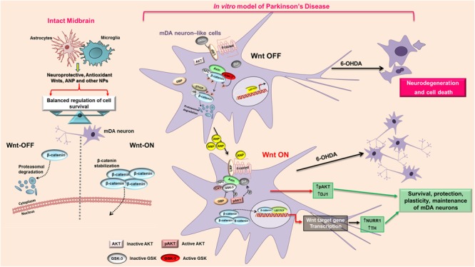
In the last decades increasing evidence indicated a crucial role of the Wnt/β-catenin signaling in development of midbrain dopaminergic (mDA) neurons. Recently dysregulation of this pathway has been proposed as a novel pathomechanism leading to Parkinson's disease (PD) and some of the molecules participating to the signaling have been evaluated as potential therapeutic targets for PD. Atrial natriuretic peptide (ANP) is a cardiac-derived hormone having a critical role in cardiovascular homeostasis. ANP and its receptors (NPRs) are widely expressed in mammalian central nervous system (CNS) where they could be implicated in the regulation of neural development, synaptic transmission and information processing, as well as in neuroprotection. Until now, the effects of ANP in the CNS have been mainly ascribed to the binding and activation of NPRs. We have previously demonstrated that ANP affects the Wnt/β-catenin signaling in colorectal cancer cells through a Frizzled receptor-mediated mechanism. The purpose of this study was to investigate if ANP is able to exert neuroprotective effect on two in vitro models of PD, and if this effect could be related to activation of the Wnt/β-catenin signaling. As cellular models of DA neurons, we used the proliferating or RA-differentiated human neuroblastoma cell line SH-SY5Y. In both DA neuron-like cultures, ANP is able to positively affect the Wnt/β-catenin signaling, by inducing β-catenin stabilization and nuclear translocation. Importantly, activation of the Wnt pathway by ANP exerts neuroprotective effect when these two cellular systems were subjected to neurotoxic insult (6-OHDA) for mimicking the neurodegeneration of PD. Our data support the relevance of exogenous ANP as an innovative therapeutic molecule for midbrain, and more in general for brain diseases for which aberrant Wnt signaling seems to be involved.

Keywords: Parkinson’s disease; Wnt/β-catenin pathway; atrial natriuretic peptide; neurodegeneration; neuroprotection.


Figures

FIGURE 1
FIGURE 1
Phenotypical characterization of proliferating SHSY5Y cells (SHSY5Ywt). (A) Confocal microscopic images showing the intracellular distribution of the neuronal markers Nestin, Tubulin-β3 and NeuN, of the DA neuron specific marker TH, and of the most important mediator of the canonical Wnt signaling β-catenin. For Tubulin-β3, Nestin, TH, and β-catenin immunostaining, cell nuclei were counterstained with propidium iodide (PI, red hue); for NeuN, cell morphology was visualized by differential interference contrast (DIC). For each marker, both single staining and merged images with PI or DIC are shown. Bars: 25 μm. (B) Western blot analysis of expression levels of Nestin, Tubulin-β3, NeuN, TH, Fzd1 receptor and β-catenin in total cell lysates from SHSY5Ywt cells.
FIGURE 2
FIGURE 2
Phenotypical characterization of RA-differentiated SHSY5Y cells (SHSY5Ydiff). (A) Resuming scheme of the protocol used for the RA-induced differentiation of SHSY5Y cells. SHSY5Ydiff cells were obtained through a 9-days differentiation process. Cells were seeded at a density of 4 × 10⁴ cells/cm2, and after 24 h (day 2), when the confluence of the monolayer was about 75%, were subjected to the 1st pulse with 10 μM RA, by replacing the culture medium with fresh medium containing RA and low serum (1% FBS). The 2nd and the 3rd pulses were performed at day 5 and 8, respectively, by replacing the medium to replenish RA, and at day 10 cells were analyzed or used for the experiments. (B) Phase contrast microscopy of SHSY5Ydiff cells 24 h after the 1st, 2nd, and 3rd pulses of 10 μM RA, showing the ongoing of neuronal differentiation with neurite outgrowth. Bar: 100 μm. (C) WB and densitometric analyses of the expression levels c-Myc and p21 in SHSY5Ydiff vs SHSY5Ywt. (D) Cytofluorimetric analysis of DNA content in SHSY5Ywt and SHSY5Ydiff cells. Values from densitometric analyses were normalized to β-actin. Significance SHSY5Ydiff vs SHSY5Ywt (Student’s t-test): ∗∗, p < 0.01; the mean ± SD; n = 4.
FIGURE 3
FIGURE 3
Comparative analyses of phenotypical features in SHSY5Ywt and SHSY5Ydiff cells. (A) Confocal microscopy showing the intracellular distribution of Tubulin-β3, NeuN, TH and β-catenin in basal and RA-differentiated SHSY5Y cultures. For Tubulin-β3, TH, and β-catenin immunostaining, cell nuclei were counterstained with propidium iodide (red hue); for NeuN, cell morphology was visualized by differential interference contrast (DIC) and merged images NeuN/DIC are shown. Bars: 25 μm. (B–D) WB and densitometric analyses of the expression levels of Tubulin-β3, NeuN and TH, Nurr1 (neuronal and DA neuron specific markers), DJ-1 and phospho-AktT308 (neuronal survival factors involved in neuroprotection against oxidative stress), and Fzd1 (Wnt pathway related molecule), in SHSY5Ywt and SHSY5Ydiff cells. Values from all densitometric analyses were normalized to β-actin or GAPDH. Significance SHSY5Ydiff vs SHSY5Ywt (Student’s t-test): p < 0.05; ∗∗, p < 0.01; the mean ± SD; n = 3.
FIGURE 4
FIGURE 4
Atrial natriuretic peptide (ANP) modifies β-catenin intracellular distribution and inhibits β-catenin degradation in SHSY5Ywt cells. (A) WB analyses of total and phosphorylated β-catenin (pβ-cateninT41/S65), and of total and phosphorylated GSK-3β (pGSK-3βSer9) levels in total cell lysates from ANP treated SHSY5Ywt cells, compared to that from untreated control and Wnt1a treated samples; analyses were performed after 3, 6, and 24 h of treatment. (B) Results from the densitometric analysis of total and phosphorylated β-catenin and GSK-3β, performed using the ImageJ processing program [http://rsbweb.nih.gov/ij/]; values were normalized to β-actin, for total β-catenin and GSK-3β, or vs the total levels of each protein for the phosphorylated forms. Results are the mean from three independent experiments. Significance vs untreated relative control: p < 0.05; ∗∗p < 0.01; the mean ± SD; n = 3. (C) Confocal microscopic images showing the intracellular distribution of β-catenin in control and ANP or Wnt1a treated cells after 6 and 24 h of culture. Bar: 25 μm. (D) Effect of ANP on the transcription of β-catenin (β-cat), LEF1 and TH genes in SHSY5Ywt cells. Cells were treated with 100 nM ANP and processed for RT-qPCR analysis after 3, 6, and 24 h of treatment. Significance vs control (Ctr): p < 0.05; ∗∗p < 0.01; the mean ± SD; n = 3.
FIGURE 5
FIGURE 5
Atrial natriuretic peptide induces β-catenin stabilization and nuclear translocation in SHSY5Ydiff cells. Confocal microscopic images showing the intracellular distribution of β-catenin (green hue) in untreated control and in ANP or Wnt1a treated cells after 24 h of culture. Cell morphology was visualized by differential interference contrast (DIC), and merged images β-catenin/DIC are also shown. White and yellow arrows point to nuclear or cytoplasmic accumulation of β-catenin, respectively. Bar: 25 μm.
FIGURE 6
FIGURE 6
Atrial natriuretic peptide pre-treatment prevents 6-OHDA toxicity on SHSY5Ywt and SHSY5Ydiff cells. (A,B) Phase contrast microscopy of SHSY5Ywt (A) and SHSY5Ydiff (B) cells exposed to 50 and 100 μM of 6-OHDA, respectively, for mimicking the neurodegeneration of PD. To verify the ability of ANP in preventing the 6-OHDA induced cytotoxicity, cells were pre-treated with 100 nM of the natriuretic peptide, 30 min or 24 h prior to 6-OHDA addition to cell culture medium. Original magnification: left panels: 10x; right panels: 20x. (C,D) Cell viability assay performed on SHSY5Ywt (C) and SHSY5Ydiff (D) cells after 24 h of 6-OHDA treatment by Trypan blue dye exclusion method; results, reported as percentage of live/dead cells, are mean of three independent experiments. Significance vs untreated control (Ctr); #Significance vs 6OH-Dopa: p < 0.05; ∗∗p < 0.01; ∗∗∗p < 0.001; the mean ± SD; n = 3.
FIGURE 7
FIGURE 7
Atrial natriuretic peptide pre-treatment reverses the neurotoxin-induced changes in β-catenin phosphorylation and degradation and in the expression of DA neuron specific markers and survival factors in SHSY5Ywt cells. WB and densitometric analysis of the expression levels of Tubulin-β3, NeuN and TH, Nurr1 (neuronal and DA neuron specific markers), DJ-1 and phospho-AktT308 (neuronal survival factors involved in neuroprotection against oxidative stress), and total and phosphorylated β-catenin (pβ-cateninT41/S65,Wnt pathway related molecules), in SHSY5Ywt cells subjected to 24 h 6-OHDA challenging in absence and in presence of 30 min or 24 h pre-treatment with ANP. Values from densitometric analysis were normalized to β-actin, or vs the total levels of β-catenin or AKT for the phosphorylated forms. Significance vs untreated control (Ctr); #Significance vs 6OH-Dopa: p < 0.05; ∗∗p < 0.01; ∗∗∗p < 0.001; the mean ± SD; n = 3.
FIGURE 8
FIGURE 8
Atrial natriuretic peptide pre-treatment reverses the neurotoxin-induced changes in β-catenin phosphorylation and degradation and in the expression of DA neuron specific markers and survival factors in SHSY5Ydiff cells. WB (A) and densitometric (B–D) analyses of the expression levels of TH, Nurr1, DJ-1 and phospho-AktT308, and total and phosphorylated β-catenin (pβ-cateninT41/S65), in SHSY5Ydiff cells subjected to 7h and 24h 6-OHDA challenging in absence and in presence of 30 min or 24 h pre-treatment with ANP. In the panel of WB (A), the duration of each treatment is reported above the corresponding lane. Values from densitometric analysis were normalized to β-actin, or vs the total levels of β-catenin or AKT for the phosphorylated forms. Significance vs untreated control (Ctr); #Significance vs 6OH-Dopa: p < 0.05; ∗∗p < 0.01; ∗∗∗p < 0.001; the mean ± SD; n = 3.
FIGURE 9
FIGURE 9
Proposed model of the mechanism that mediates the neuroprotective action of ANP on PD cellular model. In intact midbrain, the balancing between Wnt-ON and Wnt-OFF in mDA is regulated by the microglial/astrocytic component, that produce and release neuroprotective molecules including Wnts, ANP and other NPs. In the in vitro model of PD, 6-OHDA antagonizes the Wnt/β-catenin signaling and shifts the balancing toward the “Wnt OFF” state, leading to the up-regulation of active GSK-3β with a consequent phosphorylation and rapid β-catenin degradation, thus increasing cell death of mDA neuron-like cells. ANP, possibly by a direct interaction with the Frizzled receptor, activates the Wnt/β-catenin signaling cascade, induces β-catenin stabilization and nuclear translocation, up-regulates the survival factors pAKT and DJ-1 and activates transcription of the DA neuron markers Nurr1 and TH, thus leading to survival and protection of mDA neurons. APC, adenomatous polyposis coli; AKT, Serine/Threonine Protein Kinase; β-TrCP, E3 ubiquitin ligase; CK1, casein kinase 1; DKK, Dickkopf; Dvl, Disheveled; GBP, GSK3-binding protein; GSK, glycogen synthase kinase; LRP, LDL receptor-related protein; P, phosphorylation; TCF/LEF1, T-cell factor/lymphoid enhancer factor1.

Similar articles

Cited by

References

    1. Arenas E. (2014). Wnt signaling in midbrain dopaminergic neuron development and regenerative medicine for Parkinson’s disease. J. Mol. Cell Biol. 6 42–53. 10.1093/jmcb/mju001 - DOI - PubMed
    1. Berwick D. C., Harvey K. (2014). The regulation and deregulation of Wnt signaling by PARK genes in health and disease. J. Mol. Cell Biol. 6 3–12. 10.1093/jmcb/mjt037 - DOI - PMC - PubMed
    1. Bonifati V., Rizzu P., van Baren M. J., Schaap O., Breedveld G. J., Krieger E., et al. (2003). Mutations in the DJ-1 gene associated with autosomal recessive early-onset parkinsonism. Science 299 256–259. 10.1126/science.1077209 - DOI - PubMed
    1. Canet-Aviles R. M., Wilson M. A., Miller D. W., Ahmad R., McLendon C., Bandyopadhyay S., et al. (2004). The Parkinson’s disease protein DJ-1 is neuroprotective due to cysteine-sulfinic acid-driven mitochondrial localization. Proc. Natl. Acad. Sci. U.S.A. 101 9103–9108. 10.1073/pnas.0402959101 - DOI - PMC - PubMed
    1. Cao L. H., Yang X. L. (2008). Natriuretic peptides and their receptors in the central nervous system. Prog. Neurobiol. 84 234–248. 10.1016/j.pneurobio.2007.12.003 - DOI - PubMed

LinkOut - more resources