

Early detection of myocardial dysfunction using two-dimensional speckle tracking echocardiography in a young cat with hypertrophic cardiomyopathy
- PMID: 29449957
- PMCID: PMC5808971
- DOI: 10.1177/2055116918756219
Early detection of myocardial dysfunction using two-dimensional speckle tracking echocardiography in a young cat with hypertrophic cardiomyopathy
Abstract
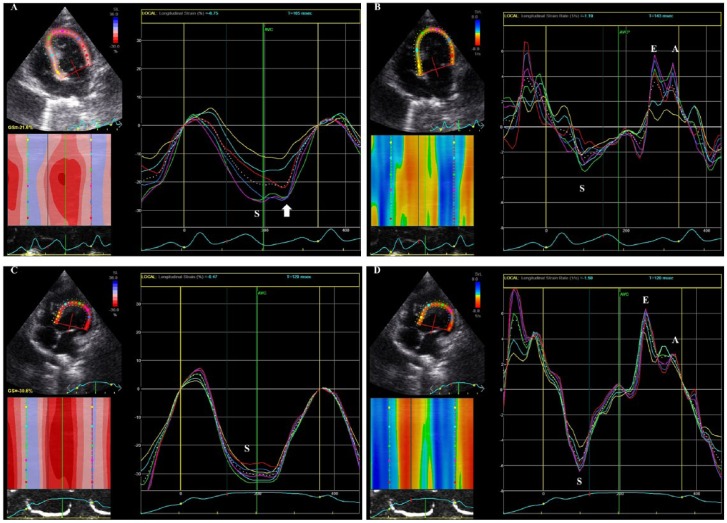
Case summary: A 5-month-old intact female Scottish Fold cat was presented for cardiac evaluation. Careful auscultation detected a slight systolic murmur (Levine I/VI). The findings of electrocardiography, thoracic radiography, non-invasive blood pressure measurements and conventional echocardiographic studies were unremarkable. However, two-dimensional speckle tracking echocardiography revealed abnormalities in myocardial deformations, including decreased early-to-late diastolic strain rate ratios in longitudinal, radial and circumferential directions, and deteriorated segmental systolic longitudinal strain. At the follow-up examinations, the cat exhibited echocardiographic left ventricular hypertrophy and was diagnosed with hypertrophic cardiomyopathy using conventional echocardiography.
Relevance and novel information: This is the first report on the use of two-dimensional speckle tracking echocardiography for the early detection of myocardial dysfunction in a cat with hypertrophic cardiomyopathy; the myocardial dysfunction was detected before the development of hypertrophy. The findings from this case suggest that two-dimensional speckle tracking echocardiography can be useful for myocardial assessment when conventional echocardiographic and Doppler findings are ambiguous.
Conflict of interest statement
Conflict of interest: The authors declared no potential conflicts of interest with respect to the research, authorship, and/or publication of this article.
Figures
Similar articles
-
Assessment of left ventricular systolic and diastolic abnormalities in patients with hypertrophic cardiomyopathy using real-time three-dimensional echocardiography and two-dimensional speckle tracking imaging.Cardiovasc Ultrasound. 2018 Oct 2;16(1):23. doi: 10.1186/s12947-018-0142-y. Cardiovasc Ultrasound. 2018. PMID: 30285887 Free PMC article.
-
Determination of multidirectional myocardial deformations in cats with hypertrophic cardiomyopathy by using two-dimensional speckle-tracking echocardiography.J Feline Med Surg. 2017 Dec;19(12):1283-1289. doi: 10.1177/1098612X17691896. Epub 2017 Feb 3. J Feline Med Surg. 2017. PMID: 28152671 Free PMC article.
-
Systolic and diastolic function assessment in fabry disease patients using speckle-tracking imaging and comparison with conventional echocardiographic measurements.J Am Soc Echocardiogr. 2013 Dec;26(12):1407-14. doi: 10.1016/j.echo.2013.09.005. Epub 2013 Oct 11. J Am Soc Echocardiogr. 2013. PMID: 24125876
-
Efficacy of echocardiography for differential diagnosis of left ventricular hypertrophy: special focus on speckle-tracking longitudinal strain.J Echocardiogr. 2021 Jun;19(2):71-79. doi: 10.1007/s12574-020-00508-3. Epub 2021 Jan 18. J Echocardiogr. 2021. PMID: 33460030 Free PMC article. Review.
-
Clinical aspects of left ventricular diastolic function assessed by Doppler echocardiography following acute myocardial infarction.Dan Med Bull. 2001 Nov;48(4):199-210. Dan Med Bull. 2001. PMID: 11767125 Review.
Cited by
-
Relationship Between Cardiac Troponin I Concentration and Myocardial Function in Hypertrophic Cardiomyopathy Cats With or Without Left Ventricular Outflow Tract Obstruction.Animals (Basel). 2025 May 1;15(9):1313. doi: 10.3390/ani15091313. Animals (Basel). 2025. PMID: 40362128 Free PMC article.
-
Assessment of myocardial function in obstructive hypertrophic cardiomyopathy cats with and without response to medical treatment by carvedilol.BMC Vet Res. 2019 Oct 28;15(1):376. doi: 10.1186/s12917-019-2141-0. BMC Vet Res. 2019. PMID: 31660967 Free PMC article.
-
Comparative study of myocardial function in cases of feline hypertrophic cardiomyopathy with and without dynamic left-ventricular outflow-tract obstruction.Front Vet Sci. 2023 Jun 22;10:1191211. doi: 10.3389/fvets.2023.1191211. eCollection 2023. Front Vet Sci. 2023. PMID: 37426078 Free PMC article.
-
Early detection of myocardial dysfunction in a cat that gradually progressed to endomyocardial form of restrictive cardiomyopathy.BMC Vet Res. 2021 Aug 14;17(1):274. doi: 10.1186/s12917-021-02987-7. BMC Vet Res. 2021. PMID: 34391430 Free PMC article.
-
Left and Right Myocardial Functionality Assessed by Two-Dimensional Speckle-Tracking Echocardiography in Cats with Restrictive Cardiomyopathy.Animals (Basel). 2021 May 28;11(6):1578. doi: 10.3390/ani11061578. Animals (Basel). 2021. PMID: 34071192 Free PMC article.
References
-
- Payne JR, Brodbelt DC, Luis Fuentes V. Cardiomyopathy prevalence in 780 apparently healthy cats in rehoming centres (the CatScan study). J Vet Cardiol 2015; 17 Suppl 1: S244–S57. - PubMed
-
- Liu Y, Deng Y, Li X, et al. Assessment of left ventricular longitudinal regional myocardial systolic function by strain imaging echocardiography in patients with hypertrophic cardiomyopathy. J Huazhong Univ Sci Technolog Med Sci 2005; 25: 703–705. - PubMed
-
- Serri K, Reant P, Lafitte M, et al. Global and regional myocardial function quantification by two-dimensional strain: application in hypertrophic cardiomyopathy. J Am Coll Cardiol 2006; 47: 1175–1181. - PubMed
Publication types
LinkOut - more resources
Full Text Sources
Other Literature Sources
Miscellaneous