

Microstructurally Anchored Cardiac Kinematics by Combining In Vivo DENSE MRI and cDTI
- PMID: 29450409
- PMCID: PMC5808941
- DOI: 10.1007/978-3-319-59448-4_36
Microstructurally Anchored Cardiac Kinematics by Combining In Vivo DENSE MRI and cDTI
Abstract
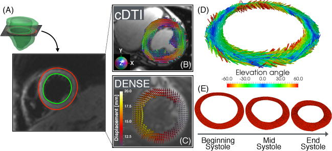
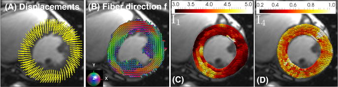
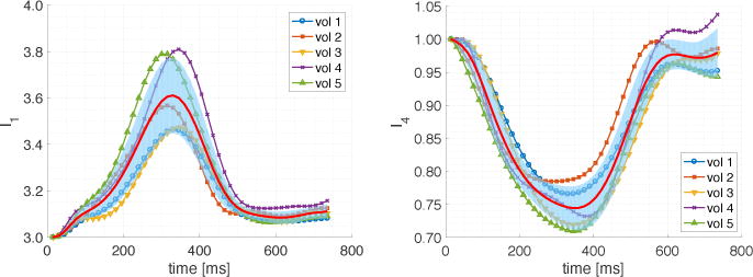
Metrics of regional myocardial function can detect the onset of cardiovascular disease, evaluate the response to therapy, and provide mechanistic insight into cardiac dysfunction. Knowledge of local myocardial microstructure is necessary to distinguish between isotropic and anisotropic contributions of local deformation and to quantify myofiber kinematics, a microstructurally anchored measure of cardiac function. Using a computational model we combine in vivo cardiac displacement and diffusion tensor data to evaluate pointwise the deformation gradient tensor and isotropic and anisotropic deformation invariants. In discussing the imaging methods and the model construction, we identify potential improvements to increase measurement accuracy. We conclude by demonstrating the applicability of our method to compute myofiber strain in five healthy volunteers.
Keywords: Cardiac deformation invariants; Cardiac kinematics; Diffusion tensor imaging; Myofiber strain.
Figures
References
-
- Aliotta E, Wu HH, Ennis DB. Convex optimized diffusion encoding (CODE) gradient waveforms for minimum echo time and bulk motion–compensated diffusion-weighted MRI. Magnetic resonance in medicine. 2016 - PubMed
-
- Ennis DB, Kindlmann G. Orthogonal tensor invariants and the analysis of diffusion tensor magnetic resonance images. Magnetic Resonance in Medicine. 2006;55(1):136–146. - PubMed
Grants and funding
LinkOut - more resources
Full Text Sources
Other Literature Sources