. 2018 Apr;9(2):173-187.
doi: 10.1007/s13244-018-0595-4. Epub 2018 Feb 15.

Major pancreatic resections: normal postoperative findings and complications

Affiliations

Major pancreatic resections: normal postoperative findings and complications

Marco Chincarini et al. Insights Imaging. 2018 Apr.

Abstract

Objectives: (1) To illustrate and describe the main types of pancreatic surgery; (2) to discuss the normal findings after pancreatic surgery; (3) to review the main complications and their radiological findings.

Background: Despite the decreased postoperative mortality, morbidity still remains high resulting in longer hospitalisations and greater costs. Imaging findings following major pancreatic resections can be broadly divided into "normal postoperative alterations" and real complications. The former should regress within a few months whereas complications may be life-threatening and should be promptly identified and treated.

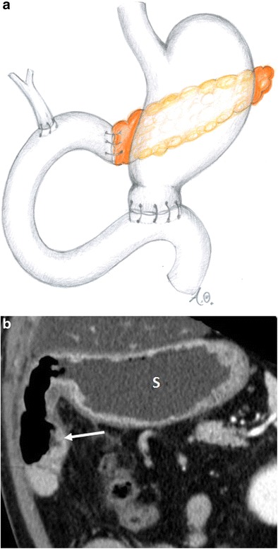
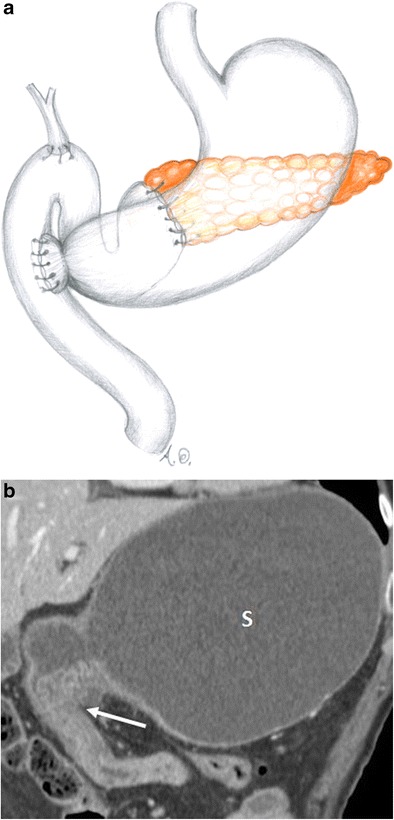
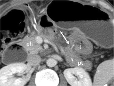
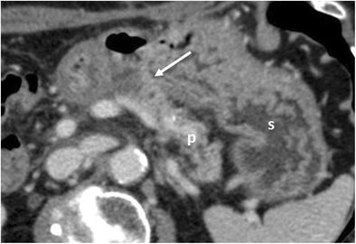
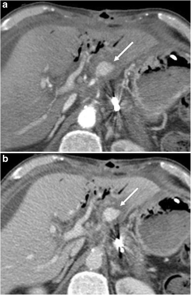
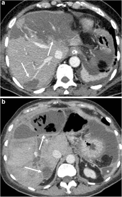
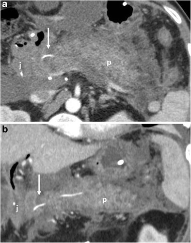
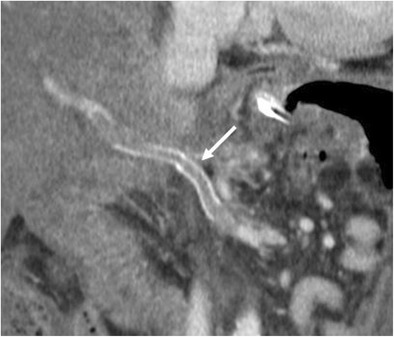
Imaging findings: CT is the most effective postoperative imaging technique. MRI and fluoroscopy are used less often and only in specific cases such as assessing the gastro-intestinal function or the biliary tree. The most common normal postoperative findings are pneumobilia, perivascular cuffing, fluid collections, lymphadenopathy, acute anastomotic oedema and stranding of the peri-pancreatic/mesenteric fat. Imaging depicts the anastomoses and the new postoperative anatomy. It can also demonstrate early and late complications: pancreatic fistula, haemorrhage, delayed gastric emptying, hepatic infarction, acute pancreatitis of the remnant, porto-mesenteric thrombosis, abscess, biliary anastomotic leaks, anastomotic stenosis and local recurrence.

Conclusions: Radiologists should be aware of surgical procedures, postoperative anatomy and normal postoperative imaging findings to better detect complications and recurrent disease.

Teaching points: • Morbidity after pancreatic resections is high. • CT is the most effective postoperative imaging technique. • Imaging depicts the anastomoses and the new postoperative anatomy. • Pancreatic fistula is the most common complication after partial pancreatic resection.

Keywords: Pancreas; Pancreatectomy; Pancreaticoduodenectomy; Pancreaticojejunostomy; Postoperative complications.


Figures

Fig. 1
Fig. 1
Whipple procedure (a, b). a Drawing. b Coronal CT image. The stomach (s) and the gastrojejunostomy (white arrow) after the Whipple procedure are visible
Fig. 2
Fig. 2
Pylorus-preserving pancreaticoduodenectomy (a, b). a Drawing. b Coronal CT image. The duodenojejunostomy (white arrow) is visible. The stomach (s) is visualised
Fig. 3
Fig. 3
Distal pancreatectomy (a, b). a Drawing. b Axial CT image. The head of the pancreas (p) is visible after a distal pancreatectomy. The resection margin (white arrow) is located at the level of the superior mesenteric vein (white arrowhead)
Fig. 4
Fig. 4
Central pancreatectomy. The head of the pancreas (ph) and the pancreatic tail (pt) are visible. The pancreaticojejunostomy to the distal pancreatic remnant (white arrow) and the anastomotic jejuna loop (j) are visible
Fig. 5
Fig. 5
Pancreaticojejunostomy. The pancreas (p), jejunal anastomotic loop (j) and pancreaticojejunostomy (white arrow) are visible after a Whipple procedure
Fig. 6
Fig. 6
Pancreaticogastrostomy. The pancreas (p), stomach (s) and pancreaticogastrostomy (white arrow) are visible after a pancreaticoduodenectomy
Fig. 7
Fig. 7
Hepaticojejunostomy (a, b). a The hepaticojejunostomy (white arrow) and jejunum (j) are visible. The common hepatic duct (white arrowhead) is slightly dilated and a small amount of aerobilia (black arrow) is visualised. b MRCP better depicts the hepaticojejunostomy (white arrow)
Fig. 8
Fig. 8
Pneumobilia and pneumowirsung (a, b). a There is a fair amount of air in the main and in left bile ducts (black arrow). The hepaticojejunostomy (white arrow) and anastomotic jejunal loop (j) are visualised. b The pancreas (p), anastomotic jejunal loop (s) and pancreaticojejunostomy (white arrow) are visualised. The main pancreatic duct is mildly dilated with an air bubble within (black arrow)
Fig. 9
Fig. 9
Perivascular cuffing. a, b) Axial CT images show a thickening (white arrows in a and b) of the fat tissue surrounding the superior mesenteric vessels in a and caeliac trunk in b. This stranding can be very focal and mass-like. In a, a postoperative reactive thickening of the anterior left pararenal fascia and a fluid collection on the same side (*) are visible
Fig. 10
Fig. 10
Postoperative collections (a, b). a Axial CT image shows a homogeneous fluid collection with a thin wall at the level of the resection margin and in the surgical bed (*) after a distal pancreatectomy. The head of the pancreas (p) is visualised. b An inhomogeneous necrotic fluid collection (*) with fat globules inside (white arrow) is visible within the left anterior pararenal fascia. These findings are consistent with a necrotic collection with steatonecrosis due to a postoperative pancreatitis after a Whipple procedure
Fig. 11
Fig. 11
Inflammatory adenopathy. Axial CT image shows the presence of enlarged lymph nodes (white arrow) in the mesentery, close to the surgical bed
Fig. 12
Fig. 12
Oedema of the pancreaticojejunostomy. Axial CT image in the immediate postoperative period shows a mild thickening of the jejunum (j) at the pancreatic anastomosis (black arrow) due to acute oedema. This condition leads to a dilation of the main pancreatic duct (white arrow). A fluid collection is visible surrounding the anastomotic jejunal loop (*)
Fig. 13
Fig. 13
Fat stranding. Coronal CT image in the immediate postoperative period shows a diffuse stranding of the mesenteric fat tissue
Fig. 14
Fig. 14
Pancreatic fistula. a, b Multiplanar CT images. The pancreatic stump (p) and jejunal loop (j) are visualised. A complete disruption of the pancreatic anastomosis is evident (black arrow in b). At the level of the anastomosis a fluid collection with multiple air bubbles inside is visible (white arrows), a finding that strongly suggests the presence of a pancreatic fistula
Fig. 15
Fig. 15
Pancreatic fistula. a Coronal curvilinear CT reconstruction shows the presence of a fluid collection (*) close to the resection margin of the pancreas (p) after a distal pancreatectomy. The presence of amylase from a surgical drainage (not shown in a) was consistent with a pancreatic fistula. b, c Spot images during ERCP show the passage of contrast material through the main pancreatic duct (white arrows) in the collection (*), a finding diagnostic for a leakage of pancreatic juice at the resection margin
Fig. 16
Fig. 16
Pancreatic fistula after the Whipple procedure. a, b Sequential images acquired during fistulography. Contrast medium is injected through a drainage on the left. Immediate filling of a fistulous tract (white arrow) and a collection (*) is seen. In the later phases passage of contrast medium inside the anastomotic loop becomes evident (black arrow in b), findings diagnostic for an anastomotic dehiscence. The main pancreatic duct is visualised (white arrowhead in b)
Fig. 17
Fig. 17
Axial CT image. A severely distended stomach filled with fluid and air is evident (s)
Fig. 18
Fig. 18
a, b Axial CT curvilinear reconstructions show an active extravasation in the arterial phase (white arrow in a) within the lumen of the jejunal anastomotic loop (j). Bleeding becomes more evident in the late phase (white arrow in b). The pancreatic stump is seen (p)
Fig. 19
Fig. 19
a, b) Axial CT images show an active extravasation in the arterial phase (white arrow in a) coming from the common hepatic artery after a Whipple procedure. Bleeding becomes more evident in the venous phase (white arrow in b)
Fig. 20
Fig. 20
Hepatic infarction. a CT image shows multiple hypodense and hypovascular areas of infarction (white arrows) following a pancreaticoduodenectomy. b CT image 4 weeks later in the same patient. The areas of infarction show a thickened and enhancing wall with multiple air bubbles within (white arrows), findings consistent with hepatic abscesses. Another abscess is visible at the level of the left lateroconal fascia (*)
Fig. 21
Fig. 21
Acute pancreatitis. a, b The pancreatic remnant (p), jejunal loop (j) and pancreatic anastomosis (white arrow) are visible. The pancreas is thickened and oedematous, surrounded by a discrete amount of fluid that extends along the anterior pararenal fascia, findings highly suggestive of an acute necrotic postoperative pancreatitis
Fig. 22
Fig. 22
Portal vein thrombosis. CT curvilinear coronal reconstruction image shows the presence of a massive thrombosis of the superior mesenteric and portal venous axis (white arrow) following a DCP with a resection of the portal vein and prosthetic reconstruction
Fig. 23
Fig. 23
Abdominal abscess. Axial CT image shows the presence of a fluid collection with a thick and enhancing wall (*) close to posterior aspect of the stomach
Fig. 24
Fig. 24
Biliary fistula. Coronal oblique CT image shows the biliary anastomosis (white arrow) surrounded by an ill-defined fluid collection (*). The jejunal loop is visible (j)
Fig. 25
Fig. 25
Spot image obtained during fistulography shows the passage of contrast medium through the biliary anastomosis inside the jejunal loop (j), a finding diagnostic for an anastomotic dehiscence. The biliary tree (white arrow) and the main pancreatic duct (black arrow) are also visible
Fig. 26
Fig. 26
Anastomotic stricture. Axial CT image shows a dilation of the main pancreatic duct associated with atrophy of the pancreatic parenchyma (white arrowhead). No signs of local tumour recurrence are seen at the pancreatic anastomosis (white arrow)
Fig. 27
Fig. 27
MRCP reveals a marked dilation of the main pancreatic duct and of some branch ducts (white arrow)
Fig. 28
Fig. 28
Tumour recurrence. Axial CT image shows the presence of hypodense soft tissue (white arrow) consistent with tumour recurrence, encasing the origin of the caeliac trunk and portal vein
Fig. 29
Fig. 29
Nodal tumour recurrence. Axial CT image shows the presence of an enlarged necrotic lymph node (white arrow) along the superior mesenteric vessels, consistent with tumour recurrence

References

    1. Whipple AO. The rationale of radical surgery for cancer of the pancreas and ampullary region. Ann Surg. 1941;114(4):612–615. doi: 10.1097/00000658-194111440-00008. - DOI - PMC - PubMed
    1. Yamauchi FI, Ortega CD, Blasbalg R, Rocha MS, Jukemura J, Cerri GG. Multidetector CT evaluation of the postoperative pancreas. Radiographics. 2012;32(3):743–764. doi: 10.1148/rg.323105121. - DOI - PubMed
    1. Morgan DE. Imaging after pancreatic surgery. Radiol Clin N Am. 2012;50(3):529–545. doi: 10.1016/j.rcl.2012.03.004. - DOI - PubMed
    1. Scialpi M, Scaglione M, Volterrani L, et al. Imaging evaluation of post pancreatic surgery. Eur J Radiol. 2005;53(3):417–424. doi: 10.1016/j.ejrad.2004.12.013. - DOI - PubMed
    1. Zamboni GA, Kruskal JB, Vollmer CM, Baptista J, Callery MP, Raptopoulos VD. Pancreatic adenocarcinoma: value of multidetector CT angiography in preoperative evaluation. Radiology. 2007;245(3):770–778. doi: 10.1148/radiol.2453061795. - DOI - PubMed

LinkOut - more resources