Review
. 2018 Apr;9(2):149-172.
doi: 10.1007/s13244-017-0591-0. Epub 2018 Feb 15.

Endometriosis: clinical features, MR imaging findings and pathologic correlation

Affiliations
Review

Endometriosis: clinical features, MR imaging findings and pathologic correlation

Pietro Valerio Foti et al. Insights Imaging. 2018 Apr.

Abstract

Objective: We illustrate the magnetic resonance imaging (MRI) features of endometriosis.

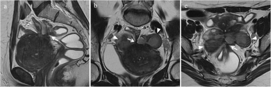
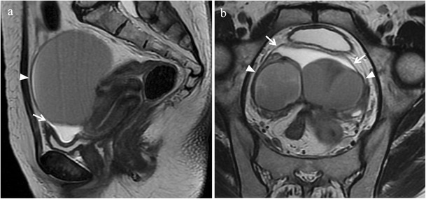
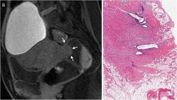
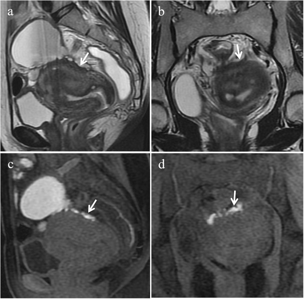
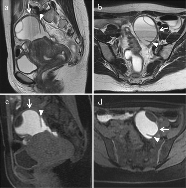
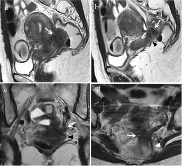
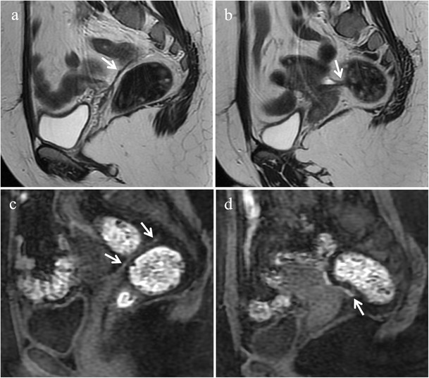
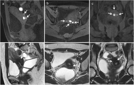
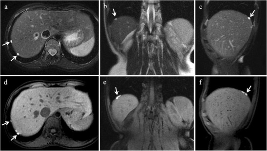
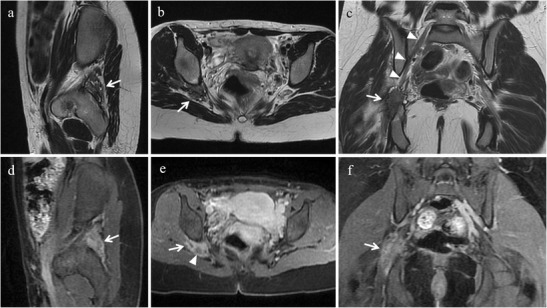
Background: Endometriosis is a chronic gynaecological condition affecting women of reproductive age and may cause pelvic pain and infertility. It is characterized by the growth of functional ectopic endometrial glands and stroma outside the uterus and includes three different manifestations: ovarian endometriomas, peritoneal implants, deep pelvic endometriosis. The primary locations are in the pelvis; extrapelvic endometriosis may rarely occur. Diagnosis requires a combination of clinical history, invasive and non-invasive techniques. The definitive diagnosis is based on laparoscopy with histological confirmation. Diagnostic imaging is necessary for treatment planning. MRI is as a second-line technique after ultrasound. The MRI appearance of endometriotic lesions is variable and depends on the quantity and age of haemorrhage, the amount of endometrial cells, stroma, smooth muscle proliferation and fibrosis. The purpose of surgery is to achieve complete resection of all endometriotic lesions in the same operation.

Conclusion: Owing to the possibility to perform a complete assessment of all pelvic compartments at one time, MRI represents the best imaging technique for preoperative staging of endometriosis, in order to choose the more appropriate surgical approach and to plan a multidisciplinary team work.

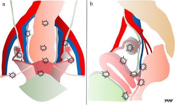
Teaching points: • Endometriosis includes ovarian endometriomas, peritoneal implants and deep pelvic endometriosis. • MRI is a second-line imaging technique after US. • Deep pelvic endometriosis is associated with chronic pelvic pain and infertility. • Endometriosis is characterized by considerable diagnostic delay. • MRI is the best imaging technique for preoperative staging of endometriosis.

Keywords: Deep infiltrating endometriosis; Endometrioma; Endometriosis; Magnetic resonance imaging; Pelvic pain; Pelvis.


Figures

Fig. 1
Fig. 1
Drawings of the female pelvis in the (a) ventral and (b) lateral views illustrate the primary locations of endometriotic lesions
Fig. 2
Fig. 2
Ureter visualization with intravenous gadolinium and variability of the ureteral course in two different patients with bilateral endometriomas. 27-year-old woman (a, b). (a) Axial contrast-enhanced fat-suppressed T1-weighted image (acquired about 10 min after contrast administration) and (b) corresponding coronal MIP image. The right ureter (white arrows) courses along the lateral margin of the right endometrial cyst (white arrowhead) and the uterus (white *), the left ureter (white arrows) courses along the posterior margin of the left endometrioma (white arrowheads). 35-year-old woman (b, c). (c) Axial contrast-enhanced fat-suppressed T1-weighted image (acquired about 10 min after contrast administration) and (d) corresponding coronal MIP image. The right ureter (white arrows) courses medially to the omolateral endometrial cyst (white arrowheads), the left ureter (white arrows) courses laterally to the omolateral endometrioma (white arrowheads). Preoperative knowledge of ureteral course is important in order to prevent iatrogenic injuries to the ureters
Fig. 3
Fig. 3
Adhesions in a 40-year-old woman with dysmenorrhea, who underwent two previous surgical interventions for endometriosis. (a) Sagittal, (b) oblique axial, and (c) oblique coronal T2-weighted images show spiculated hypointense strands arranged in confluent angles (white arrows) with loss of the cleavage planes among the anterior surface of the sigma, the posterior uterine serosa and bilateral endometriomas (white arrowheads)
Fig. 4
Fig. 4
Adhesions in a 43-year-old woman. (a) Sagittal, and (b) oblique axial T2-weighted images display spiculated hypointense strands (white arrows) between the anterior surface of the sigma and the posterior uterine serosa with angulation of rectosigmoid colon
Fig. 5
Fig. 5
Indirect sign of adhesions in a 36-year-old woman with bilateral endometriomas. (a) Sagittal and (b) oblique coronal T2-weighted images show free fluid in the pelvis on “anti-declive position” (white arrows), due to the presence of adhesions and bilateral endometriomas (white arrowheads). Both ovaries are joined together involved in adhesions (kissing ovaries)
Fig. 6
Fig. 6
Deep infiltrating endometriosis in a 34-year-old woman. (a) Sagittal fat-suppressed T1-weighted image shows an endometriotic nodule (white arrows) infiltrating the muscular layer of the anterior rectal wall. The lesion displays homogeneous intermediate signal intensity due to fibrous tissue and smooth muscle. (b) Photomicrograph (H&E 250X). Focus of endometriosis without haemorrhage
Fig. 7
Fig. 7
Deep infiltrating endometriosis in a 48-year-old woman. (a) Sagittal fat-suppressed T1-weighted image displays an endometriotic nodule (white arrows) infiltrating the muscular layer of the anterior rectal wall. Within the lesion hyperintense foci are detectable (white arrowheads), representing hemorrhagic content. (b) Photomicrograph (H&E 200×). Focus of endometriosis with marked haemorrhage. On (c) sagittal T2-weighted image the solid endometriotic lesion appears mainly hypointense (white arrows), with small hyperintense cystic foci inside representing dilated ectopic endometrial glands (white arrowheads)
Fig. 8
Fig. 8
Bladder endometriosis with extrinsic involvement in a 21-year-old woman with left endometrioma and other endometriotic implants involving the left fallopian tube, the uterine serosa and the right uterosacral ligament. (a) Coronal and (b) sagittal fat-suppressed T1-weighted images demonstrate one spot of high signal intensity on the serosal surface of the bladder representing a small peritoneal implant with hemorrhagic content (white arrows). Note the endometrioma in the left ovary (white arrowhead in a). On (c) sagittal T2-weighted image the implant (white arrow) is hardly detectable because it is partly masked by the hypointense signal of the bladder wall
Fig. 9
Fig. 9
Bladder endometriosis with intrinsic involvement in a 35-year-old woman with dysuria, hematuria and urinary incontinence during menses. (a) Sagittal and (b) oblique axial T2-weighted images show a mural mass with low signal intensity infiltrating the posterior bladder wall (white arrows). On (c) axial T2* image punctate signal voids due to hemosiderin deposition can be seen along the borders of the lesion (white arrow). On (d) sagittal and (e) oblique axial fat-suppressed T1-weighted images the implant displays intermediate signal intensity with spots of high signal intensity, representing hemorrhagic content (white arrows). (f) Oblique axial MIP image (acquired about 10 min after contrast administration) demonstrates the relationship between the lesion and the ureters (white arrowheads)
Fig. 10
Fig. 10
Ureteral endometriosis in a 35-year-old woman with multiple DIE lesions. (a) Sagittal and (b) coronal T2-weighted images show a retroperitoneal solid nodule with spiculated margins (white arrows), with low signal intensity, adjacent to left iliac vessels. (c) Sagittal and (d) coronal T2-weighted images demonstrate dilatation of the ureter upstream and of the contralateral ureter (white arrowheads). On (e) sagittal, (f) coronal and (g) axial contrast-enhanced fat-suppressed T1-weighted images the lesion displays enhancement (white arrows)
Fig. 11
Fig. 11
Ovarian endometriotic cysts in a 36-year-old woman. The same patient as in Fig. 5. (a, b) Sagittal, (c) oblique axial and (d) oblique coronal T2-weighted images show bilateral endometriomas with intermediate to low signal intensity (white arrows). The ovaries are joined together behind the uterus (kissing ovaries). Note the low signal intensity in the declivous portion of the left cyst (“shading” sign, white arrowhead in c). On (e, f) sagittal and (g) oblique axial fat-suppressed T1-weighted images the cysts demonstrate high signal intensity (white arrows). (h) Photomicrograph (H&E 20X). Ovarian endometriotic cyst
Fig. 12
Fig. 12
Endometriosis of the uterine serosa in a 46-year-old woman, who underwent a previous surgical intervention for endometrioma of the right ovary. (a) Sagittal and (b) oblique axial T2-weighted images show endometriotic implants involving the uterine serosa. The lesions demonstrate indistinct margins and low signal intensity (white arrows). On (c) sagittal and (d) coronal fat-suppressed T1-weighted images the lesions display high signal intensity (white arrows)
Fig. 13
Fig. 13
Endometriosis of the round ligaments in a 45-year-old woman with dysmenorrhea, urinary symptoms and chronic pelvic pain, who underwent a previous surgical intervention for endometrioma of the left ovary. (a-c) Oblique coronal and (d, e) oblique axial T2-weighted images. Both round ligaments (white arrows) appear thickened, nodular and shortened. (f) Oblique coronal fat-suppressed T1-weighted image reveals small intralesional high signal-intensity foci within the right ligament (white arrowheads) that represent hemorrhagic component
Fig. 14
Fig. 14
Hematosalpinx in a 46-year-old woman with endometriosis. The same patient as in Fig. 12. (a) Sagittal and (b) axial T2-weighted images show a tortuous, tubular structure with internal fluid-fluid level in the left adnexa (white arrows). On (c) sagittal and (d) axial fat-suppressed T1-weighted images endoluminal content displays high signal intensity (white arrows), a finding consistent with hematosalpinx. Note the incomplete mucosal and submucosal plicae along the tubal wall (white arrowheads)
Fig. 15
Fig. 15
Endometriosis of the retrocervical region in a 39-year-old woman with dysmenorrhea, dyspareunia, tenesmus, catamenial dyschezia and dysuria. (a, b) Sagittal, (c) oblique coronal and (d) oblique axial T2-weighted images show a retrocervical hypointense endometriotic implant (white arrows) infiltrating the posterior uterine serosa and the anterior rectal wall, extending to the left vaginal fornix (black arrowheads in b and c) and to the left uterosacral ligament (white arrowhead in d)
Fig. 16
Fig. 16
Endometriosis of the retrocervical region in a 41-year-old woman with dysmenorrhea and dyspareunia, who underwent previous laparoscopic adhesiolysis. (a) Sagittal and (b) oblique axial T2-weighted images show a hypointense ill-defined infiltrative tissue (white arrows) involving the posterior portion of the cervix, the retrocervical region and the anterior rectal wall. Small cystic cavities are seen within the lesion. On (c) sagittal and (d) axial fat-suppressed T1-weighted images the lesion exhibits intermediate signal intensity (white arrows) with small hyperintense foci reflecting haemorrhage. Note the small left hemorrhagic cyst (white arrowheads in b and d) showing a fluid-fluid level on T2-weighted image; it disappeared in the follow-up US examinations and was likely a hemorrhagic follicular cyst
Fig. 17
Fig. 17
Endometriosis of the uterosacral ligaments in a 35-year-old woman with multiple DIE lesions. The same patient as in Fig. 10. (a, b) Sagittal T2-weighted images show hypointense thickening of both uterosacral ligaments (white arrows). (c, d) Sagittal fat-suppressed T1-weighted images demonstrate hyperintense spots within the ligaments (white arrows), representing hemorrhagic foci
Fig. 18
Fig. 18
Endometriosis of the right uterosacral ligament in a 28-year-old woman with dyspareunia. (a) Oblique axial, (b) sagittal and (c) oblique coronal T2-weighted images show a low-signal-intensity endometriotic lesion with spiculated margins involving the right uterosacral ligament (white arrows). The lesion extends to the iliococcygeus muscle (white arrowheads). (d) Photomicrograph (H&E 250X). Endometriosis in the uterosacral ligament
Fig. 19
Fig. 19
Endometriosis of the vagina and cervix in a 46-year-old woman. (a) Sagittal, (b) oblique axial, (c) oblique coronal fat-suppressed T1-weighted images, and (d) sagittal, (e) oblique axial and (f) oblique coronal T2-weighted images show small endometriotic implants involving the posterior vaginal fornix and the cervix, with multiloculated appearance (white arrows) characterized by cystic areas with hyperintense content, better depicted on fat-suppressed T1-weighted images due to subacute blood products
Fig. 20
Fig. 20
Endometriosis of the anterior rectal wall in a 48-year-old woman with dysmenorrhea, chronic pelvic pain and catamenial diarrhoea. (a) Sagittal and (b) oblique coronal T2-weighted images, and (c) sagittal and (d) oblique coronal contrast-enhanced fat-suppressed T1-weighted images show an endometriotic nodule (white arrows) infiltrating the muscular layer of the anterior rectal wall. The lesion has a “fan shaped” configuration with the base adhering to the rectal wall and the apex oriented toward the retrocervical region. The implant demonstrates isointense signal compared to muscle on T2-weighted and T1-weighted sequences; the slightly high signal at the luminal side (white arrowheads in a and b) corresponds to (sub)mucosal thickening and enhances after intravenous administration of contrast material (white arrowheads in c and d). (e) Photomicrograph (H&E 40X). Endometriosis in the muscularis propria (black arrows)
Fig. 21
Fig. 21
Endometriosis of the rectal wall in a 45-year-old woman with cyclic hematochezia, constipation, pencil-like stools and episodes of intestinal subocclusion, who underwent previous right ureteral stenting. (a) Sagittal and (b) oblique coronal T2-weighted images, and (c) sagittal and (d) oblique coronal contrast-enhanced fat-suppressed T1-weighted images show an endometriotic lesion (white arrows) infiltrating the muscular and submucosal layers of the rectal wall. The lesion extends longitudinally for about 7 cm and determines severe stenosis. Note the hyperintense signal of the (sub)mucosal layer protruding into the rectal lumen (black arrowheads in a and b). (e) Photomicrograph (H&E 25X). Endometriosis in submucosa (black *) and in muscularis propria (white *)
Fig. 22
Fig. 22
Diaphragmatic endometriosis in a 28-year-old woman with right-sided basithoracic chest pain associated with menses, who underwent a previous surgical intervention for endometrioma of the left ovary. (a) Axial, (b) coronal, (c) sagittal T2-weighted images, and (d) axial, (e) coronal and (f) sagittal fat-suppressed T1-weighted images show right-sided hyperintense nodular diaphragmatic implants (white arrows). The lesions are better depicted on fat-suppressed T1-weighted sequences
Fig. 23
Fig. 23
Extrapelvic sciatic nerve endometriosis in a 31-year-old woman who has been suffering from cyclic sciatica for about 2 years. (a) Sagittal, (b) axial and (c) coronal T2-weighted images show hypointense spiculated soft-tissue thickening centred around the right sciatic nerve at the sciatic notch (white arrows). (d) Sagittal, (e) axial and (f) coronal contrast-enhanced fat-suppressed T1-weighted images display enhancement of the mass (white arrows). Note the sciatic nerve cephalad to the lesion (white arrowheads in c) and within the lesion (white arrowhead in e)
Fig. 24
Fig. 24
Indirect MR findings of sciatic nerve endometriosis. The same patient as in Fig. 23. (a) Axial and (b) coronal T2-weighted images show atrophy of right obturator internus (white arrows) and gemellus superior muscle (white arrowhead in a) compared with the contralateral ones (white arrows and white arrowhead in a). (c) Axial and (d) coronal T2-weighted images display atrophy of right piriformis muscle compared with the contralateral one (white arrows)

References

    1. Bazot M, Bharwani N, Huchon C, et al. European society of urogenital radiology (ESUR) guidelines: MR imaging of pelvic endometriosis. Eur Radiol. 2017;27(7):2765–2775. doi: 10.1007/s00330-016-4673-z. - DOI - PMC - PubMed
    1. Coutinho A, Jr, Bittencourt LK, Pires CE, et al. MR imaging in deep pelvic endometriosis: a pictorial essay. Radiographics. 2011;31(2):549–567. doi: 10.1148/rg.312105144. - DOI - PubMed
    1. Chamié LP, Blasbalg R, Pereira RM, Warmbrand G, Serafini PC. Findings of pelvic endometriosis at transvaginal US, MR imaging, and laparoscopy. Radiographics. 2011;31(4):E77–100. doi: 10.1148/rg.314105193. - DOI - PubMed
    1. Adamson GD, Kennedy SH, Hummelshoj L. Creating solutions in endometriosis: global collaboration through the world endometriosis research foundation. J Endometriosis. 2010;2:3–6.
    1. Di Paola V, Manfredi R, Castelli F, Negrelli R, Mehrabi S, Pozzi Mucelli R. Detection and localization of deep endometriosis by means of MRI and correlation with the ENZIAN score. Eur J Radiol. 2015;84(4):568–574. doi: 10.1016/j.ejrad.2014.12.017. - DOI - PubMed

LinkOut - more resources