

Nurr1 promotes neurogenesis of dopaminergic neuron and represses inflammatory factors in the transwell coculture system of neural stem cells and microglia
- PMID: 29450981
- PMCID: PMC6489950
- DOI: 10.1111/cns.12825
Nurr1 promotes neurogenesis of dopaminergic neuron and represses inflammatory factors in the transwell coculture system of neural stem cells and microglia
Abstract
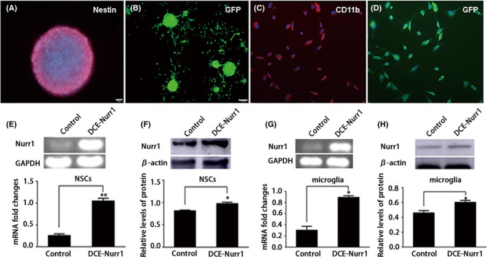
Introduction: Neural stem cells (NSCs) are the most promising cells for cell replacement therapy for Parkinson's disease (PD). However, a majority of the transplanted NSCs differentiated into glial cells, thereby limiting the clinical application. Previous studies indicated that chronic neuroinflammation plays a vital role in the degeneration of midbrain DA (mDA) neurons, which suggested the developing potential of therapies for PD by targeting the inflammatory processes. Thus, Nurr1 (nuclear receptor-related factor 1), a transcription factor, has been referred to play a pivotal role in both the differentiation of dopaminergic neurons in embryonic stages and the maintenance of the dopaminergic phenotype throughout life.
Aim: This study investigated the effect of Nurr1 on neuroinflammation and differentiation of NSCs cocultured with primary microglia in the transwell coculture system.
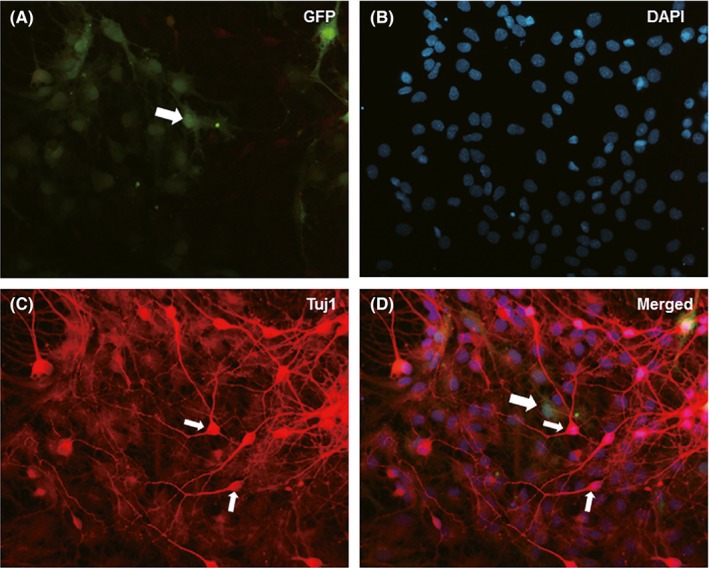
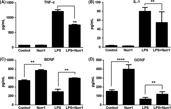
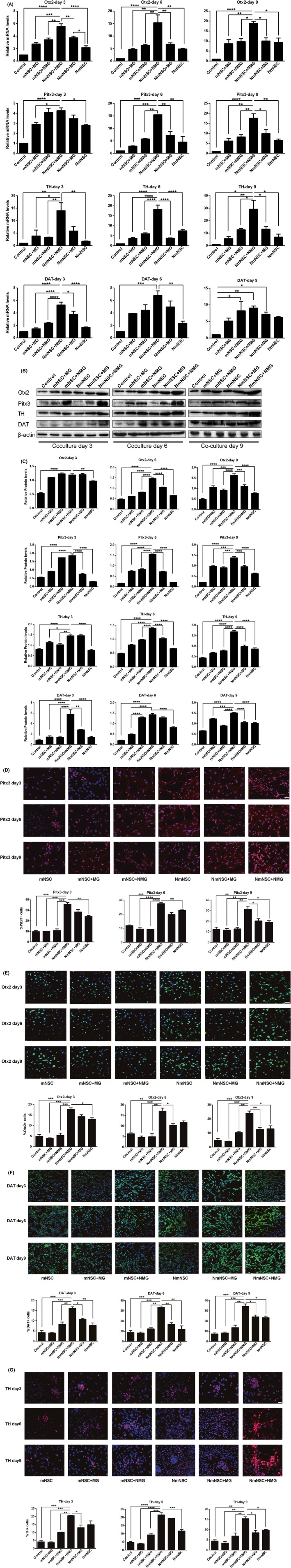
Results: The results showed that Nurr1 exerted anti-inflammatory effects and promoted the differentiation of NSCs into dopaminergic neurons.
Conclusions: The results suggested that Nurr1 protects dopaminergic neurons from neuroinflammation insults by limiting the production of neurotoxic mediators by microglia and maintain the survival of transplanted NSCs. These phenomena provided a new theoretical and experimental foundation for the transplantation of Nurr1-overexpressed NSCs as a potential treatment of PD.
Keywords: Nurr1; coculture; inflammatory; microglia; neural stem cells.
© 2018 John Wiley & Sons Ltd.
Figures
Similar articles
-
Transplantation of Nurr1-overexpressing neural stem cells and microglia for treating parkinsonian rats.CNS Neurosci Ther. 2020 Jan;26(1):55-65. doi: 10.1111/cns.13149. Epub 2019 May 13. CNS Neurosci Ther. 2020. PMID: 31087449 Free PMC article.
-
The co-transduction of Nurr1 and Brn4 genes induces the differentiation of neural stem cells into dopaminergic neurons.Cell Biol Int. 2011 Dec;35(12):1217-23. doi: 10.1042/CBI20110028. Cell Biol Int. 2011. PMID: 21663595
-
Role of Nurr1 in the Generation and Differentiation of Dopaminergic Neurons from Stem Cells.Neurotox Res. 2016 Jul;30(1):14-31. doi: 10.1007/s12640-015-9586-0. Epub 2015 Dec 17. Neurotox Res. 2016. PMID: 26678495
-
Advances in NURR1-Regulated Neuroinflammation Associated with Parkinson's Disease.Int J Mol Sci. 2022 Dec 19;23(24):16184. doi: 10.3390/ijms232416184. Int J Mol Sci. 2022. PMID: 36555826 Free PMC article. Review.
-
The function and mechanisms of Nurr1 action in midbrain dopaminergic neurons, from development and maintenance to survival.Int Rev Neurobiol. 2012;102:1-22. doi: 10.1016/B978-0-12-386986-9.00001-6. Int Rev Neurobiol. 2012. PMID: 22748824 Review.
Cited by
-
Zinc finger E-Box binding homeobox 2 (ZEB2)-induced astrogliosis protected neuron from pyroptosis in cerebral ischemia and reperfusion injury.Bioengineered. 2021 Dec;12(2):12917-12930. doi: 10.1080/21655979.2021.2012551. Bioengineered. 2021. PMID: 34852714 Free PMC article.
-
Potent synthetic and endogenous ligands for the adopted orphan nuclear receptor Nurr1.Exp Mol Med. 2021 Jan;53(1):19-29. doi: 10.1038/s12276-021-00555-5. Epub 2021 Jan 21. Exp Mol Med. 2021. PMID: 33479411 Free PMC article. Review.
-
Neuroprotective effects of a lead compound from coral via modulation of the orphan nuclear receptor Nurr1.CNS Neurosci Ther. 2023 Mar;29(3):893-906. doi: 10.1111/cns.14025. Epub 2022 Nov 23. CNS Neurosci Ther. 2023. PMID: 36419251 Free PMC article.
-
NR4A2 Exacerbates Cerebral Ischemic Brain Injury via Modulating microRNA-652/Mul1 Pathway.Neuropsychiatr Dis Treat. 2020 Oct 6;16:2285-2296. doi: 10.2147/NDT.S265601. eCollection 2020. Neuropsychiatr Dis Treat. 2020. PMID: 33116527 Free PMC article.
-
Roles of Transcription Factors in the Development and Reprogramming of the Dopaminergic Neurons.Int J Mol Sci. 2022 Jan 13;23(2):845. doi: 10.3390/ijms23020845. Int J Mol Sci. 2022. PMID: 35055043 Free PMC article. Review.
References
-
- Kalia LV, Lang AE. Parkinson's disease. Lancet. 2015;386:896‐912. - PubMed
-
- Hirsch EC, Vyas S, Hunot S. Neuroinflammation in Parkinson's disease. Parkinsonism Relat Disord. 2012;18(Suppl 1):S210‐S212. - PubMed
-
- Ransohoff RM. How neuroinflammation contributes to neurodegeneration. Science. 2016;353:777‐783. - PubMed
-
- Wirdefeldt K, Adami HO, Cole P, Trichopoulos D, Mandel J. Epidemiology and etiology of Parkinson's disease: a review of the evidence. Eur J Epidemiol. 2011;26(Suppl 1):S1‐S58. - PubMed
-
- LeWitt PA, Fahn S. Levodopa therapy for Parkinson disease: A look backward and forward. Neurology. 2016;86(14 Suppl 1):S3‐S12. - PubMed
Publication types
MeSH terms
Substances
Associated data
- Actions
LinkOut - more resources
Full Text Sources
Other Literature Sources
