

doi: 10.4103/eus.eus_49_17.
EUS elastography: How to do it?
Affiliations
- PMID: 29451165
- PMCID: PMC5838723
- DOI: 10.4103/eus.eus_49_17
Item in Clipboard
EUS elastography: How to do it?
Endosc Ultrasound.
2018 Jan-Feb.
Abstract
Strain elastography as used in EUS (EUS-real-time tissue elastography [RTE]) is a qualitative technique and provides information on the relative stiffness between one tissue and another. This article reviews the principles, technique, and interpretation of EUS-RTE in various organs. It includes information on how to optimize the technique as well as a discussion on pitfalls and artifacts. We also refer to the article describing RTE using conventional ultrasound transducers.
Keywords: EUS; real-time tissue elastography; ultrasound.
Conflict of interest statement
There are no conflicts of interest.
Figures
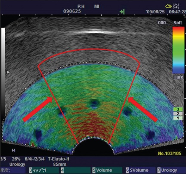
Reducing region of interest sector size to improve uniformity. Using a tightly curved array transducer to image this tissue-mimicking phantom, the material in front of the transducer face has been stressed more that the lateral margins resulting in a greater strain (red color) at the center of the sector, and less strain (blue color) induced at the lateral margins. Narrowing the sector size of the elastography region of interest (indicated by the red lines) will improve uniformity of the stress field. Using the smaller sector region of interest, the probe can be moved to interrogate the mid-portion and two lateral sector regions separately. Depth-dependent stress attenuation can also be observed in the central part in this phantom with homogeneous elasticity
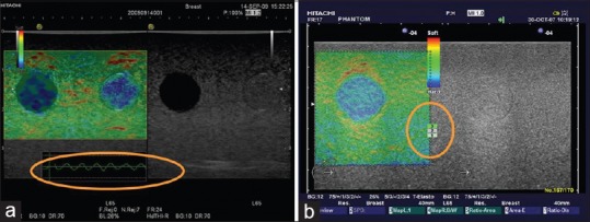
Quality parameters. (a) Strain graph display scale should be set to between 0.5% and 1.0%. (b) Press indicator: A value of 3 or 4 is recommended, but this feedback is now replaced by the strain graph display in recent versions of the software
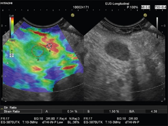
Strain ratio measurement as applied to the pancreas. Region of interest A is placed within the pancreatic mass and region of interest B in adjacent parenchyma or fat layer. Tissue-lesion ratio = B/A shown in a small solid and malignant pancreatic tumor
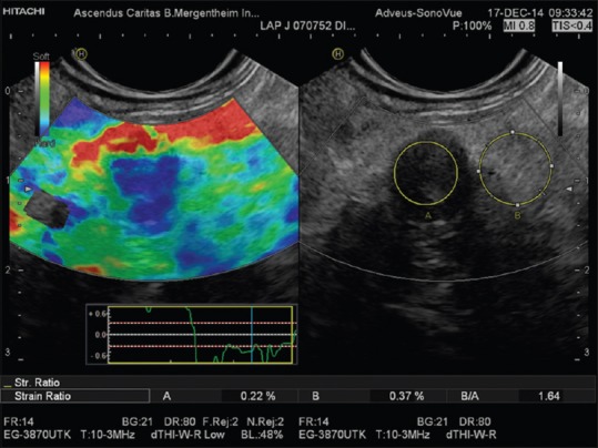
Real-time tissue elastography with strain ratio measurement as applied to the liver. Region of interest A is placed within the hyperechoic liver mass and region of interest B in adjacent liver parenchyma. The Strain ratio of 1.3 indicates nearly equal elasticity of the lesion (which turned out to be focal fatty infiltration) and surrounding “normal” liver tissue. Due to respiration-induced movements, both slipping boundaries between liver and peritoneal space show a high strain (red lines, arrows) and should not be used as a reference area for strain ratio measurements
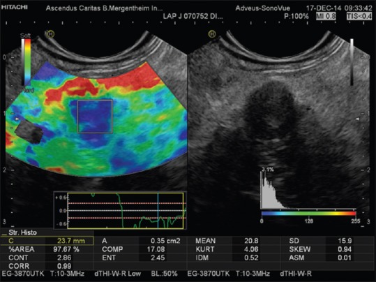
Histograms display the distribution of recorded strains (%) within an elastography region of interest of a small solid and malignant pancreatic tumor [same lesion as in Figure 3]
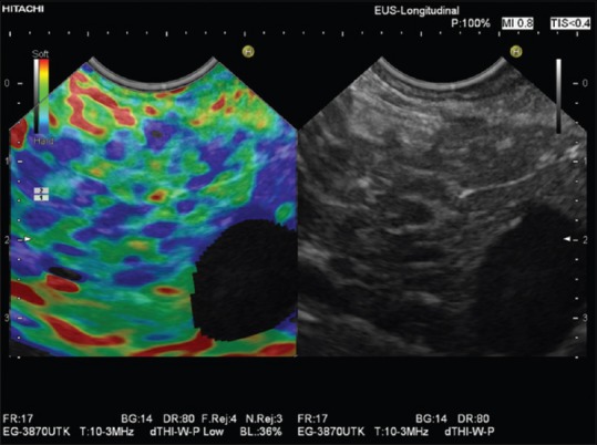
Elastographic honeycomb pattern in a patient with early chronic pancreatitis
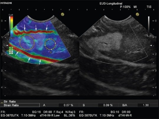
Reactive periduodenal lymph node (region of interest A). In comparison to the softest area available in the vicinity of the lymph node (region of interest B), a strain ratio of 4.4 was calculated
References
-
- Shiina T, Nightingale KR, Palmeri ML, et al. WFUMB guidelines and recommendations for clinical use of ultrasound elastography: Part 1: Basic principles and terminology. Ultrasound Med Biol. 2015;41:1126–47. - PubMed
-
- Dietrich CF, Cantisani V. Current status and perspectives of elastography. Eur J Radiol. 2014;83:403–4. - PubMed
-
- Dietrich CF. [Real Time Elastography. Indications not only in the gastrointestional tract] Endo heute. 2010;23:177–225.
-
- Dietrich CF. Multiple clinical applications. Multiple clinical solutions. Endheu. 2012;24:177–225.
-
- Bilgen M, Insana MF. Error analysis in acoustic elastography. II. Strain estimation and SNR analysis. J Acoust Soc Am. 1997;101:1147–54. - PubMed
LinkOut - more resources
Full Text Sources
Other Literature Sources
