

Conundrums and confusions regarding how polyethylene glycol-fusion produces excellent behavioral recovery after peripheral nerve injuries
- PMID: 29451204
- PMCID: PMC5840989
- DOI: 10.4103/1673-5374.224363
Conundrums and confusions regarding how polyethylene glycol-fusion produces excellent behavioral recovery after peripheral nerve injuries
Abstract
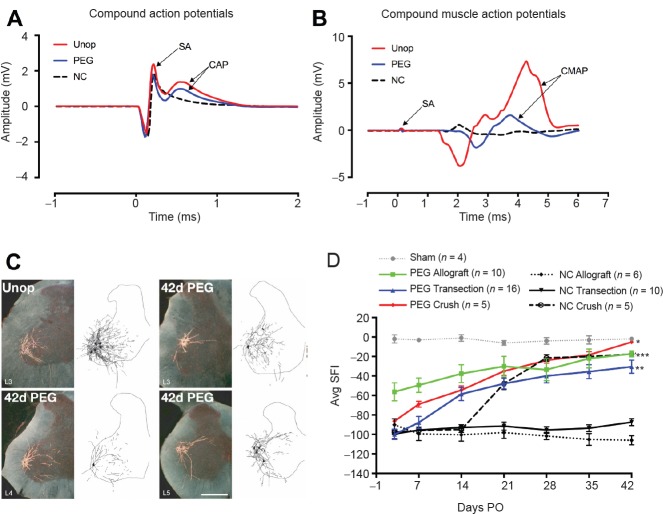
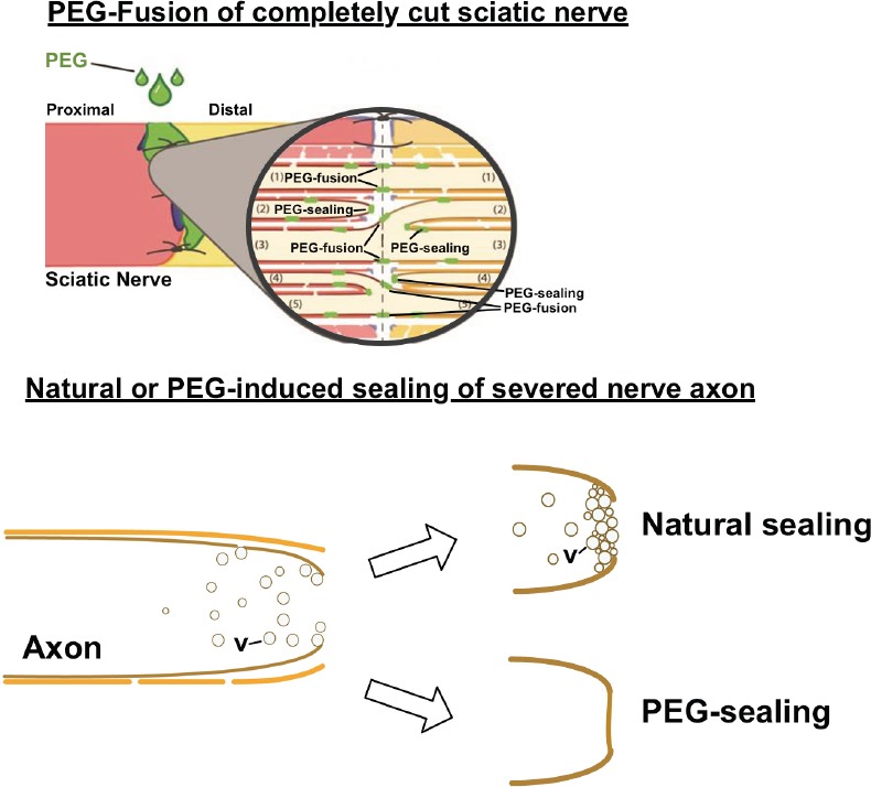
Current Neuroscience dogma holds that transections or ablations of a segment of peripheral nerves produce: (1) Immediate loss of axonal continuity, sensory signaling, and motor control; (2) Wallerian rapid (1-3 days) degeneration of severed distal axons, muscle atrophy, and poor behavioral recovery after many months (if ever, after ablations) by slowly-regenerating (1 mm/d), proximal-stump outgrowths that must specifically reinnervate denervated targets; (3) Poor acceptance of microsutured nerve allografts, even if tissue-matched and immune-suppressed. Repair of transections/ablations by neurorrhaphy and well-specified-sequences of PEG-fusion solutions (one containing polyethylene glycol, PEG) successfully address these problems. However, conundrums and confusions regarding unorthodox and dramatic results of PEG-fusion repair in animal model systems often lead to misunderstandings. For example, (1) Axonal continuity and signaling is re-established within minutes by non-specifically PEG-fusing (connecting) severed motor and sensory axons across each lesion site, but remarkable behavioral recovery to near-unoperated levels takes several weeks; (2) Many distal stumps of inappropriately-reconnected, PEG-fused axons do not ever (Wallerian) degenerate and continuously innervate muscle fibers that undergo much less atrophy than otherwise-denervated muscle fibers; (3) Host rats do not reject PEG-fused donor nerve allografts in a non-immuno-privileged environment with no tissue matching or immunosuppression; (4) PEG fuses apposed open axonal ends or seals each shut (thereby preventing PEG-fusion), depending on the experimental protocol; (5) PEG-fusion protocols produce similar results in animal model systems and early human case studies. Hence, iconoclastic PEG-fusion data appropriately understood might provoke a re-thinking of some Neuroscience dogma and a paradigm shift in clinical treatment of peripheral nerve injuries.
Keywords: Wallerian degeneration; allograft; autograft; axonal repair; axotomy; nerve regeneration; polyethylene glycol.
Conflict of interest statement
None declared.
Figures
Similar articles
-
Polyethylene glycol treated allografts not tissue matched nor immunosuppressed rapidly repair sciatic nerve gaps, maintain neuromuscular functions, and restore voluntary behaviors in female rats.J Neurosci Res. 2018 Jul;96(7):1243-1264. doi: 10.1002/jnr.24227. Epub 2018 Apr 16. J Neurosci Res. 2018. PMID: 29659046 Free PMC article.
-
Polyethylene glycol solutions rapidly restore and maintain axonal continuity, neuromuscular structures, and behaviors lost after sciatic nerve transections in female rats.J Neurosci Res. 2018 Jul;96(7):1223-1242. doi: 10.1002/jnr.24225. Epub 2018 Apr 16. J Neurosci Res. 2018. PMID: 29659058 Free PMC article.
-
Polyethylene glycol (PEG) and other bioactive solutions with neurorrhaphy for rapid and dramatic repair of peripheral nerve lesions by PEG-fusion.J Neurosci Methods. 2019 Feb 15;314:1-12. doi: 10.1016/j.jneumeth.2018.12.015. Epub 2018 Dec 23. J Neurosci Methods. 2019. PMID: 30586569 Free PMC article.
-
The curious ability of polyethylene glycol fusion technologies to restore lost behaviors after nerve severance.J Neurosci Res. 2016 Mar;94(3):207-30. doi: 10.1002/jnr.23685. Epub 2015 Nov 3. J Neurosci Res. 2016. PMID: 26525605 Free PMC article. Review.
-
Typical and atypical properties of peripheral nerve allografts enable novel strategies to repair segmental-loss injuries.J Neuroinflammation. 2022 Feb 28;19(1):60. doi: 10.1186/s12974-022-02395-0. J Neuroinflammation. 2022. PMID: 35227261 Free PMC article. Review.
Cited by
-
Interpretation of Data from Translational Rodent Nerve Injury and Repair Models.Hand Clin. 2024 Aug;40(3):429-440. doi: 10.1016/j.hcl.2024.03.004. Epub 2024 May 19. Hand Clin. 2024. PMID: 38972687 Free PMC article. Review.
-
Methylene blue enhances polyethylene glycol-fusion repair of completely severed rat sciatic nerves.Neural Regen Res. 2021 Oct;16(10):2056-2063. doi: 10.4103/1673-5374.308099. Neural Regen Res. 2021. PMID: 33642394 Free PMC article.
-
An analysis of differential gene expression in peripheral nerve and muscle utilizing RNA sequencing after polyethylene glycol nerve fusion in a rat sciatic nerve injury model.PLoS One. 2024 Sep 4;19(9):e0304773. doi: 10.1371/journal.pone.0304773. eCollection 2024. PLoS One. 2024. PMID: 39231134 Free PMC article.
-
Polyethylene glycol treated allografts not tissue matched nor immunosuppressed rapidly repair sciatic nerve gaps, maintain neuromuscular functions, and restore voluntary behaviors in female rats.J Neurosci Res. 2018 Jul;96(7):1243-1264. doi: 10.1002/jnr.24227. Epub 2018 Apr 16. J Neurosci Res. 2018. PMID: 29659046 Free PMC article.
-
Tyrosine-derived polymeric surfactant nanospheres insert cholesterol in cell membranes.J Colloid Interface Sci. 2023 Aug 15;644:264-274. doi: 10.1016/j.jcis.2023.04.045. Epub 2023 Apr 15. J Colloid Interface Sci. 2023. PMID: 37120875 Free PMC article.
References
-
- Bittner GD, Keating CP, Kane JR, Britt JM, Spaeth CS, Fan JD, Zuzek A, Wilcott RW, Thayer WP, Winograd JM, Gonzalez-Lima F, Schallert T. Rapid, effective, and long-lasting behavioral recovery produced by microsutures, methylene blue, and polyethylene glycol after completely cutting rat sciatic nerves. J Neurosci Res. 2012;90:967–980. - PubMed
-
- Brushart TM. Nerve Repair. New York: Oxford University Press; 2011.
-
- Ghergherehchi CL, Bittner GD, Hastings RL, Mikesh M, Riley DC, Trevino RC, Schallert T, Thayer WP, Sunkesula SR, Ha TA, Munoz N, Pyarali M, Bansal A, Poon AD, Mazal AT, Smith TA, Wong NS, Dunne PJ. Effects of extracellular calcium and surgical techniques on restoration of axonal continuity by polyethylene glycol fusion following complete cut or crush severance of rat sciatic nerves. J Neurosci Res. 2016;94:231–245. - PMC - PubMed
Publication types
Grants and funding
LinkOut - more resources
Full Text Sources
Other Literature Sources
