

Is it necessary to use the entire root as a donor when transferring contralateral C7 nerve to repair median nerve?
- PMID: 29451212
- PMCID: PMC5840998
- DOI: 10.4103/1673-5374.224376
Is it necessary to use the entire root as a donor when transferring contralateral C7 nerve to repair median nerve?
Abstract
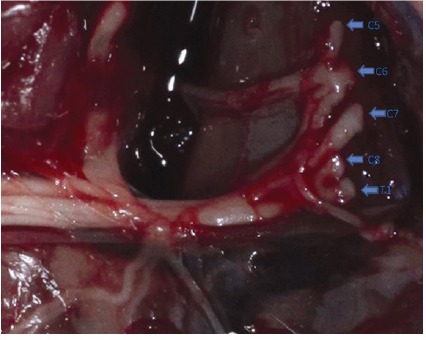
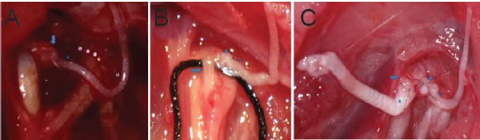
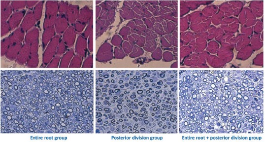
If a partial contralateral C7 nerve is transferred to a recipient injured nerve, results are not satisfactory. However, if an entire contralateral C7 nerve is used to repair two nerves, both recipient nerves show good recovery. These findings seem contradictory, as the above two methods use the same donor nerve, only the cutting method of the contralateral C7 nerve is different. To verify whether this can actually result in different repair effects, we divided rats with right total brachial plexus injury into three groups. In the entire root group, the entire contralateral C7 root was transected and transferred to the median nerve of the affected limb. In the posterior division group, only the posterior division of the contralateral C7 root was transected and transferred to the median nerve. In the entire root + posterior division group, the entire contralateral C7 root was transected but only the posterior division was transferred to the median nerve. After neurectomy, the median nerve was repaired on the affected side in the three groups. At 8, 12, and 16 weeks postoperatively, electrophysiological examination showed that maximum amplitude, latency, muscle tetanic contraction force, and muscle fiber cross-sectional area of the flexor digitorum superficialis muscle were significantly better in the entire root and entire root + posterior division groups than in the posterior division group. No significant difference was found between the entire root and entire root + posterior division groups. Counts of myelinated axons in the median nerve were greater in the entire root group than in the entire root + posterior division group, which were greater than the posterior division group. We conclude that for the same recipient nerve, harvesting of the entire contralateral C7 root achieved significantly better recovery than partial harvesting, even if only part of the entire root was used for transfer. This result indicates that the entire root should be used as a donor when transferring contralateral C7 nerve.
Keywords: animal experiment; avulsion injury; brachial plexus injury; contralateral C7 transfer; entire root; median nerve; nerve regeneration; nerve root; neural regeneration; partial root; peripheral nerve injury; ulnar nerve.
Conflict of interest statement
None declared.
Figures
Similar articles
-
Repair of brachial plexus lesions by end-to-side side-to-side grafting neurorrhaphy: experience based on 11 cases.Microsurgery. 2005;25(2):126-46. doi: 10.1002/micr.20036. Microsurgery. 2005. PMID: 15389968
-
Total brachial plexus injury: contralateral C7 root transfer to the lower trunk versus the median nerve.Neural Regen Res. 2018 Nov;13(11):1968-1973. doi: 10.4103/1673-5374.239444. Neural Regen Res. 2018. PMID: 30233071 Free PMC article.
-
The effectiveness of contralateral C7 nerve root transfer for the repair of avulsed C7 nerve root in total brachial plexus injury: an experimental study in rats.J Reconstr Microsurg. 2013 Jun;29(5):325-30. doi: 10.1055/s-0033-1343498. Epub 2013 Apr 18. J Reconstr Microsurg. 2013. PMID: 23599214
-
An electroencephalography-based human-machine interface combined with contralateral C7 transfer in the treatment of brachial plexus injury.Neural Regen Res. 2022 Dec;17(12):2600-2605. doi: 10.4103/1673-5374.335838. Neural Regen Res. 2022. PMID: 35662188 Free PMC article. Review.
-
Nerve transfer helps repair brachial plexus injury by increasing cerebral cortical plasticity.Neural Regen Res. 2014 Dec 1;9(23):2111-4. doi: 10.4103/1673-5374.147939. Neural Regen Res. 2014. PMID: 25657729 Free PMC article. Review.
Cited by
-
Modified contralateral C7 nerve transfer: the possibility of permitting ulnar nerve recovery is confirmed by 10 cases of autopsy.Neural Regen Res. 2019 Aug;14(8):1449-1454. doi: 10.4103/1673-5374.253530. Neural Regen Res. 2019. PMID: 30964072 Free PMC article.
-
Why It Is Necessary to Use the Entire Root rather than Partial Root When Doing Contralateral C7 Nerve Transfer: Cortical Plasticity Also Matters besides the Amount of Nerve Fibers.Neural Plast. 2021 Jan 4;2021:8819380. doi: 10.1155/2021/8819380. eCollection 2021. Neural Plast. 2021. PMID: 33488696 Free PMC article.
-
The Median Nerve Injury Model in Pre-clinical Research - A Critical Review on Benefits and Limitations.Front Cell Neurosci. 2019 Jun 28;13:288. doi: 10.3389/fncel.2019.00288. eCollection 2019. Front Cell Neurosci. 2019. PMID: 31316355 Free PMC article. Review.
-
Cadaveric study to assess the feasibility of S1 neurectomy and contralateral S1 transfer for spastic hemiparesis.J Orthop. 2022 Apr 21;31:99-102. doi: 10.1016/j.jor.2022.04.010. eCollection 2022 May-Jun. J Orthop. 2022. PMID: 35514531 Free PMC article.
-
Restoration of intrinsic hand function by superficial radial nerve: an anatomical study.BMC Musculoskelet Disord. 2023 Aug 2;24(1):628. doi: 10.1186/s12891-023-06758-3. BMC Musculoskelet Disord. 2023. PMID: 37532990 Free PMC article.
References
-
- Chen L, Gu YD, Hu SN, Xu JG, Xu L, Fu Y. Contralateral C7 transfer for the treatment of brachial plexus root avulsions in children: a report of 12 cases. J Hand Surg (Am) 2007;32:96–103. - PubMed
-
- Chuang DC. Neurotization procedures for brachial plexus injuries. Hand Clin. 1995;11:633–645. - PubMed
-
- Chuang DC, Cheng SL, Wei FC, Wu CL, Ho YS. Clinical evaluation of C7 spinal nerve transection: 21 patients with at least 2 years' follow-up. Br J Plast Surg. 1998;51:285–290. - PubMed
-
- Chuang DC, Hernon C. Minimum 4-year follow-up on contralateral C7 nerve transfers for brachial plexus injuries. J Hand Surg Am. 2012;37:270–276. - PubMed
LinkOut - more resources
Full Text Sources
Other Literature Sources
Miscellaneous
