. 2018 May 1;596(9):1747-1776.
doi: 10.1113/JP275228. Epub 2018 Mar 26.

Supraspinal modulation of neuronal synchronization by nociceptive stimulation induces an enduring reorganization of dorsal horn neuronal connectivity

Affiliations

Supraspinal modulation of neuronal synchronization by nociceptive stimulation induces an enduring reorganization of dorsal horn neuronal connectivity

E Contreras-Hernández et al. J Physiol. .

Abstract

Key points: The state of central sensitization induced by the intradermic injection of capsaicin leads to structured (non-random) changes in functional connectivity between dorsal horn neuronal populations distributed along the spinal lumbar segments in anaesthetized cats. The capsaicin-induced changes in neuronal connectivity and the concurrent increase in secondary hyperalgesia are transiently reversed by the systemic administration of small doses of lidocaine, a clinically effective procedure to treat neuropathic pain. The effects of both capsaicin and lidocaine are greatly attenuated in spinalized preparations, showing that supraspinal influences play a significant role in the shaping of nociceptive-induced changes in dorsal horn functional neuronal connectivity. We conclude that changes in functional connectivity between segmental populations of dorsal horn neurones induced by capsaicin and lidocaine result from a cooperative adaptive interaction between supraspinal and spinal neuronal networks, a process that may have a relevant role in the pathogenesis of chronic pain and analgesia.

Abstract: Despite a profusion of information on the molecular and cellular mechanisms involved in the central sensitization produced by intense nociceptive stimulation, the changes in the patterns of functional connectivity between spinal neurones associated with the development of secondary hyperalgesia and allodynia remain largely unknown. Here we show that the state of central sensitization produced by the intradermal injection of capsaicin is associated with structured transformations in neuronal synchronization that lead to an enduring reorganization of the functional connectivity within a segmentally distributed ensemble of dorsal horn neurones. These changes are transiently reversed by the systemic administration of small doses of lidocaine, a clinically effective procedure to treat neuropathic pain. Lidocaine also reduces the capsaicin-induced facilitation of the spinal responses evoked by weak mechanical stimulation of the skin in the region of secondary but not primary hyperalgesia. The effects of both intradermic capsaicin and systemic lidocaine on the segmental correlation and coherence between ongoing cord dorsum potentials and on the responses evoked by tactile stimulation in the region of secondary hyperalgesia are greatly attenuated in spinalized preparations, showing that supraspinal influences are involved in the reorganization of the nociceptive-induced structured patterns of dorsal horn neuronal connectivity. We conclude that the structured reorganization of the functional connectivity between the dorsal horn neurones induced by capsaicin nociceptive stimulation results from cooperative interactions between supraspinal and spinal networks, a process that may have a relevant role in the shaping of the spinal state in the pathogenesis of chronic pain and analgesia.

Keywords: analgesia; capsaicin; lidocaine; nociception; spinal cord.


Figures

Figure 1
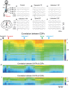
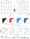
Figure 1. Systemic lidocaine reverses the capsaicin‐induced increase in correlation between ongoing spinal cord activity
AF, CDPs recorded from the L5 caudal and the L6 rostral segments on both sides and IFPs recorded at two different depths in the L6cL segment before and after capsaicin, lidocaine and spinalization, as indicated. Negativity is upward for CDPs and downward for the IFPs. The histological section on the left shows the intraspinal location of the IFP recording sites. G, changes produced by capsaicin, lidocaine and spinalization in the correlation between the paired sets of CDPs recorded with the ensemble of 12 electrodes placed along the L4–L7 segments on both sides of the spinal cord. The whole set of coefficients of correlation obtained during the 10 min Control 0 recording period is displayed in descending order as a vertical column. The coefficients of correlation obtained from 10 min non‐overlapping recordings made at subsequent times are displayed keeping the same order as the Control 0 coefficients. Colours show magnitude of correlation (see scale). Arrows show time of capsaicin and lidocaine injections and of spinalization. H and I, equivalent displays of the coefficients of correlation of the S‐IFPs and D‐IFPs with the CDPs recorded from different segments, as indicated. See text for further explanations.
Figure 2
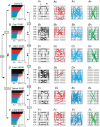
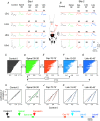
Figure 2. The patterns of segmental correlation between CDPs are disrupted after the intradermic injection of capsaicin and temporarily restored by systemic lidocaine
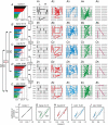
A, horizontal display of the coefficients of correlation obtained from all the combinations between paired sets of the CDPs recorded during the control period ordered according to their magnitude and separated in four different ranges as shown by colours. A1–A4, spinal cord diagrams showing the segmental location of the paired sets of CDPs used to calculate the coefficients of correlation in each range. Lines indicate segmental location of CDP recording sites. B, correlograms and B1–B4, segmental distribution of coefficients obtained from recordings made 70–80 min after the injection of capsaicin. Note in panel B1 increased correlation between CDPs recorded from neighbouring segments. CC4, the effects of capsaicin are reversed 10–20 min after the systemic injection of lidocaine. DD4, restoration of the effects of capsaicin 80–90 min after the injection of lidocaine. EE4, spinalization removes the post‐lidocaine increase in correlation. FF4, after a second injection of lidocaine the segmental distribution of the coefficients of correlation resembles the configuration attained 10–20 min after the first administration of lidocaine. The coefficients of similarity (RMSS) between correlograms generated under different experimental conditions are indicated by the brackets. Red numbers denote correlograms with highest similarity. Same experiment as that of Fig. 1. Further explanations in text.
Figure 3
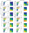
Figure 3. Differential effects of capsaicin and lidocaine on the correlation of superficial and deep intraspinal fields with the CDPs recorded from different segments
The graphs with the horizontal bars display the coefficients of correlation arranged in descending order. The segmental distribution of these coefficients is shown in the right. In both graphs the colours indicate the magnitude of the correlation (see scale). Separate plots were made for the correlations of the S‐IFPs and D‐IFPs with the CDPs as indicated. Location of intraspinal electrodes is shown in Fig. 1. The brackets show the RMSS values between different pairs of correlograms. Numbers in red indicate the lowest RMSS values, suggesting similar distributions. Same experiment as that of Figs 1 and 2. See text for further explanations.
Figure 4
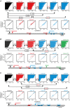
Figure 4. The differential effects of capsaicin on the functional connectivity between dorsal horn neurones are transiently reversed by lidocaine and suppressed by spinalization
Panels AI show the graphs obtained by plotting the control coefficients of correlation between paired sets of CDPs (Control 0, abscissae) versus the coefficients obtained at different times before and during the action of capsaicin (AC), after lidocaine (DG), after spinalization (H) and after a second administration of lidocaine (I). Note that after capsaicin the coefficients of correlation were separated in two distinct clusters that persisted without substantial changes until the injection of lidocaine transiently reversed the effects of capsaicin giving rise to a single cluster. After spinalization the post‐lidocaine two‐cluster arrangement of the coefficients changed to a single cluster. The RMSS similarity coefficients between the different correlograms as well as the ANCOVA P s values for the C1 and C2 clusters are included in the figure. JR , effects of capsaicin, lidocaine and spinalization on the correlation of the S‐IFPS and D‐IFPs with the CDPs. Data obtained from the same experiment as that of Figs 1, 2, 3. See text for further details.
Figure 5
Figure 5. Consistency of effects on correlation between CDPs produced by capsaicin and lidocaine in preparations with intact neuraxis
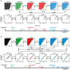
A–C, data from three different experiments showing correlograms and graphs relating control coefficients of correlation versus effects produced by capsaicin and lidocaine as indicated. Note that despite the differences in the control correlograms in the three experiments, capsaicin increased the correlation between CDPs and lidocaine transiently reversed the effects of capsaicin. RSMM coefficients of similarity between different correlograms are indicated in the figure. Bars at the bottom show timing of the different procedures. See text for further details.
Figure 6
Figure 6. Systemic lidocaine transiently reverses the capsaicin‐induced increase in power spectra and coherence between CDPs
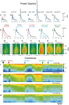
AC, power spectra of the CDPs recorded from segments L6cL (black traces) and L6cR (blue traces) before, 10–20 min and 80–90 min after the intradermic injection of capsaicin. D and E power spectra obtained from recordings made 10–20 min and 80–90 min after the systemic administration of lidocaine. F, 10–20 min after spinalization. G, second dose of lidocaine injected 60–70 min after spinalization. HL , superposed traces of the normalized spectra of the L6cL CDPs allow comparison of the changes in the different frequency components produced by capsaicin, lidocaine and spinalization, as indicated (see colours). MS, segmental distribution of the changes in power spectra produced by capsaicin, lidocaine and spinalization. Graphs show frequency of power spectra versus segmental location of the recording sites. Frequency changes in left (L) and right (R) sides are plotted separately as mirror images (see abscissa). The colours indicate the magnitude of the power spectra in logarithmic scale (see calibration). Note the expansion of the capsaicin‐induced spectral increase towards the more rostral segments and the transient suppression of this effect by lidocaine. TW, changes in coherence between CDPs produced by the different experimental procedures in four frequency ranges as indicated (see red arrows and grey bars in control spectra displayed in A). Note that the capsaicin increase in coherence is largest in the low frequency range (1.5–4.5 Hz). Same experiment as that of Figs 1 and 2. Further explanations in text.
Figure 7
Figure 7. Supraspinal dependence of the effects of capsaicin and lidocaine on the correlation between ongoing CDPs and IFPs
Same format as that of Fig. 1. AE, raw recordings of the CDPs and IFPs obtained before and after spinalization, capsaicin and lidocaine, as indicated. F, vertical display of the coefficients of correlation obtained from sets of 5 min continuous recordings displayed, taking as reference the distribution of the Control 0 coefficients. G and H, correlation of S‐IFPs and D‐IFPs with CDPs. Diagram shows spinal location of IFP recording sites. See text for further explanations.
Figure 8
Figure 8. The effects of capsaicin and lidocaine on the segmental distribution of the correlation between the CDPs are subject to supraspinal control
A–E, same format as that of Fig. 2. The effects of the different procedures are indicated in each panel. Note that after spinalization the segmental distribution of the coefficients of correlation was not significantly changed by capsaicin and lidocaine. The RMSS values between different correlograms are indicated. FJ, graphs obtained by plotting the control coefficients of correlation between CDPs (Control 0, abscissae) versus the coefficients obtained at different times as indicated. P s > 0.05 for both C1 and C2 in Spinal 10–15 min vs. Cap 65–70 min, Cap 65–70 min vs. Lido 15–20 min and Lido 15–20 min vs. Lido 55–60 min. See text for further explanations.
Figure 9
Figure 9. Changes in correlation produced by capsaicin and lidocaine in previously spinalized preparations
A and B, data from two different experiments showing correlograms and graphs relating control coefficients of correlation versus changes induced by different procedures as indicated. Same format as that of Fig. 5. Note that after spinalization, capsaicin and lidocaine had rather small effects on the correlation between CDPs. RMSS values between different correlograms, best linear fits and P s values are indicated in the figures. Bars at the bottom show timing of the different procedures. See text for further details.
Figure 10
Figure 10. Spinalization greatly attenuates the effects of capsaicin and lidocaine on the power spectra and coherence between CDPs seen in preparations with intact neuroaxis
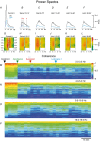
Same format as that of Fig. 6. AF, changes in the power spectra of CDPs recorded from segments L6rL (black traces) and L6rR (blue traces) during several experimental procedures, as indicated. G–L, graphs showing frequency versus segmental location of the changes in power spectra produced by spinalization, capsaicin and lidocaine. Note that after spinalization, capsaicin slightly increased the power spectra in the low frequency range and that this effect was mildly reduced by lidocaine, particularly on the right side. Recordings of L7rR were not available. MP, changes in coherence between CDPs produced by the different experimental procedures in four frequency ranges as indicated. Note that lidocaine had a rather weak action on the capsaicin changes induced after spinalization, particularly for frequencies above 9.5 Hz. Further explanations in text.
Figure 11
Figure 11. Systemic lidocaine transiently reverses the facilitation of the spinal responses evoked by mechanical stimulation in the region of secondary hyperalgesia as well as the capsaicin‐induced disruption of correlation between CDPs
A, CDPs produced by mechanical stimulation of the skin with an air puff applied close to the site of capsaicin injection (Site 1). B, same as A, following mechanical stimulation farther away from the capsaicin‐injection site (35 mm), within the region of secondary hyperalgesia (Site 2). The numbers indicate percentage changes in peak amplitude of the mechanically evoked responses relative to the amplitude of the control responses. C–F, changes in the coefficients of correlation between paired sets of CDPs produced by capsaicin and lidocaine at the indicated times. Numbers show the RMSS values between pairs of correlograms obtained at different times after capsaicin and lidocaine, as indicated. GJ, plots of the Control 0 coefficients (abscissae) against the correlation coefficients obtained under the different experimental procedures (ordinates). The graphs H and J show that the separation between the two clusters observed 60–70 min after capsaicin was transiently reduced 30–40 min after lidocaine. At that time the correlogram resembled the control one (RMSS value 0.34). 50–60 min after lidocaine the coefficients were again distributed in two similar clusters resembling those displayed 60–70 min after capsaicin (P > 0.05 for both C1 and C2). Bar at the bottom shows timing of the different procedures. See text for further details.
Figure 12
Figure 12. Acute spinalization strongly attenuates the effects of capsaicin and lidocaine on the responses produced by mechanical stimulation of the skin as well as on the correlation between CDPs
Same format as Fig. 11. A, effects of spinalization, capsaicin and lidocaine on the CDPs recorded in the rostral and caudal regions of the L5 and L6 segments following tactile stimulation of the skin close to the site of capsaicin injection (Site 1, primary hyperalgesia). B, effects on CDPs evoked by mechanical stimulation away from the capsaicin‐injection site (35 mm), within the region of secondary hyperalgesia (Site 2). The numbers indicate percentage changes in peak amplitude of the mechanically evoked responses relative to the amplitude of the responses produced after spinalization. CG, changes in the coefficients of correlation between CDPs produced by capsaicin and lidocaine at the indicated times. RMSS values between correlograms are shown. HL, plots of the Control 0 coefficients (abscissae) against the correlation coefficients obtained under different experimental procedures (ordinates). Note that spinalization separated the coefficients into two clusters. Capsaicin slightly reduced the correlation between the paired sets of CDPs grouped in cluster C2, with practically no effect on the correlation between CDPs in cluster C1. Lines show best linear fits. P s < 0.05 for C2 and P s > 0.05 for C1 in Spinal 30–35 min vs. Cap 70–75 min. P s > 0.05 for C1 and C2 in Cap 70–75 min vs. Lido 15–20 min and Lido 15–20 min vs. Lido 40–45 min. Bar at the bottom shows timing of the different procedures. See text for further explanations.

Similar articles

Cited by

References

    1. Abaei M, Sagar DR, Stockley EG, Spicer CH, Prior M, Chapman V & Auer DP (2016). Neural correlates of hyperalgesia in the monosodium iodoacetate model of osteo‐arthritis pain. Mol Pain 12, 1744806916642445. - PMC - PubMed
    1. Abarbanel HDI, Rabinovich MI, Selverston A, Bazhenov MV, Huerta R, Sushchik MM & Rubchinskii LL (1996). Synchronization in neural networks. Physics‐Uspekhi 39, 337–362.
    1. Averbeck BB & Lee D (2004). Coding and transmission of information by neural ensembles. Trends Neurosci 27, 225–230. - PubMed
    1. Bak P (1997). How Nature Works: The Science of Self‐organized Criticality. Oxford University Press, Oxford, UK.
    1. Basbaum A, Bautista DM, Scherrer G & Julius D (2009). Cellular and molecular mechanisms of pain. Cell 139, 267–284. - PMC - PubMed

Publication types

LinkOut - more resources