. 2019 Jan;28(1):41-55.
doi: 10.1002/pro.3390. Epub 2018 Mar 10.

Can dimedone be used to study selenoproteins? An investigation into the reactivity of dimedone toward oxidized forms of selenocysteine

Affiliations

Can dimedone be used to study selenoproteins? An investigation into the reactivity of dimedone toward oxidized forms of selenocysteine

N Connor Payne et al. Protein Sci. 2019 Jan.

Abstract

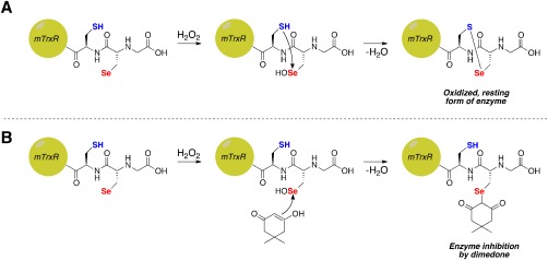
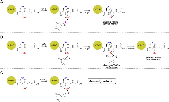
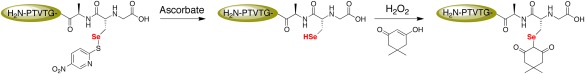
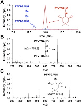
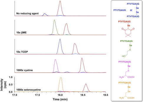
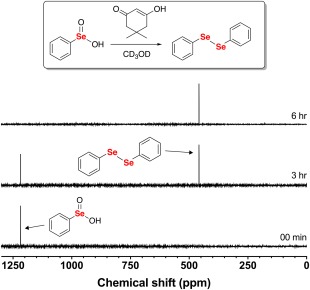
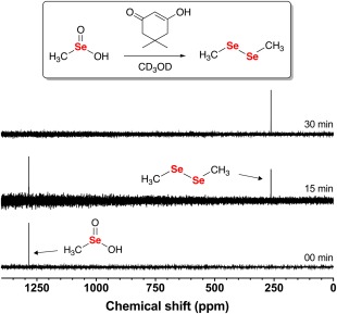
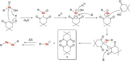
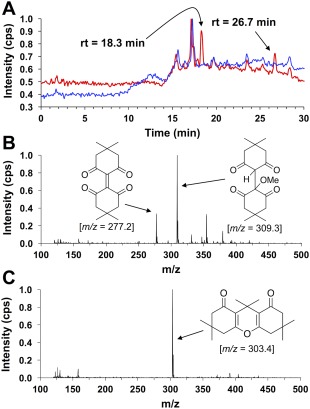
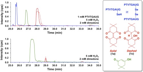
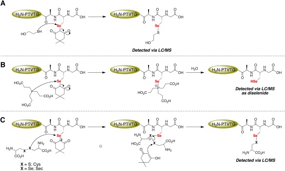
Dimedone is a widely used reagent to assess the redox state of cysteine-containing proteins as it will alkylate sulfenic acid residues, but not sulfinic acid residues. While it has been reported that dimedone can label selenenic acid residues in selenoproteins, we investigated the stability, and reversibility of this label in a model peptide system. We also wondered whether dimedone could be used to detect seleninic acid residues. We used benzenesulfinic acid, benzeneseleninic acid, and model selenocysteine-containing peptides to investigate possible reactions with dimedone. These peptides were incubated with H2 O2 in the presence of dimedone and then the reactions were followed by liquid chromatography/electrospray ionization mass spectrometry (LC/ESI-MS). The native peptide, H-PTVTGCUG-OH (corresponding to the native amino acid sequence of the C-terminus of mammalian thioredoxin reductase), could not be alkylated by dimedone, but could be carboxymethylated with iodoacetic acid. However the "mutant peptide," H-PTVTGAUG-OH, could be labeled with dimedone at low concentrations of H2 O2 , but the reaction was reversible by addition of thiol. Due to the reversible nature of this alkylation, we conclude that dimedone is not a good reagent for detecting selenenic acids in selenoproteins. At high concentrations of H2 O2 , selenium was eliminated from the peptide and a dimeric form of dimedone could be detected using LCMS and 1 H NMR. The dimeric dimedone product forms as a result of a seleno-Pummerer reaction with Sec-seleninic acid. Overall our results show that the reaction of dimedone with oxidized cysteine residues is quite different from the same reaction with oxidized selenocysteine residues.

Keywords: Pummerer reaction; dimedone; selenenic acid; seleninic acid; selenocysteine; sulfenic acid; sulfinic acid.


Figures

Figure 1
Figure 1
Possible fates of Sec‐selenenic acid in mTrxR. (A) Oxidation of Sec in mTrxR by H2O2 to form Sec‐selenenic acid, followed by resolution by the adjacent Cys residue resulting in the formation of a selenosulfide bond and concomitant enzyme recovery from oxidation. (B) Theoretical inhibition of mTrxR by dimedone via alkylation of Sec‐selenenic acid.
Figure 2
Figure 2
Possible reasons for the absence of inhibition of thioredoxin reductase by dimedone in the presence of H2O2. (A) Rapid resolution of Sec‐selenenic acid by the adjacent Cys residue prevents alkylation. (B) Alkylation of Sec‐selenenic acid by dimedone, followed by removal of the dimedone label (pK a ∼ 5)9 by attack of the adjacent Cys residue. (C) Over oxidation of Sec to Sec‐seleninic acid, whose reactivity toward dimedone is heretofore unknown.
Figure 3
Figure 3
Alkylation of model Sec‐containing peptide with dimedone. Sec was initially protected as Sec(5‐Npys) to prevent oxidation of the selenium atom. Ascorbate selectively removes 5‐Npys protecting group from Sec,17 generating a free Sec‐selenol, which was then oxidized by H2O2 in the presence of dimedone to alkylate the Sec‐selenenic acid.
Figure 4
Figure 4
(A) Extracted ion chromatogram monitoring the elution of species with m/z values of 751.8 (blue) or 891.3 (red) after exposing peptide H‐PTVTGAUG‐OH to alkylation conditions of 5 μM H‐PTVTGAUG‐OH, 5 μM H2O2, and 50 μM dimedone for 25 min in 100 mM potassium phosphate buffer, pH 7.0. These m/z values correspond to the H‐PTVTGAUG‐OH peptide as the diselenide and dimedone‐labeled H‐PTVTGAUG‐OH peptide, respectively. It is clear that, under these alkylation conditions, the Sec‐containing peptide H‐PTVTGAUG‐OH is oxidized to the selenenic acid and subsequently alkylated with dimedone. (B) Mass spectrum of the analyte at rt = 17.65 min. In the mass spectrum, the most intense ion is the PTVTGAUG diselenide peptide at m/z = 751.8. (C) Mass spectrum of the analyte at rt = 18.00 min. In the mass spectrum, the most intense ion is the dimedone‐labeled PTVTGAUG peptide at m/z = 891.3.
Figure 5
Figure 5
Extracted ion chromatograms monitoring the elution of species with m/z values of 751.8 and 752.8 (blue), 891.3 (red), 829.3 (green), 871.3 (magenta), and 920.2 (tangerine) after exposing dimedone‐labeled peptide H‐PTVTGAUG‐OH to conditions of (top to bottom): no reducing agent, 10× βME, 10× TCEP, 1000× cystine, or 1000× selenocystine for 15 min in 100 mM potassium phosphate buffer, pH 7.0. These m/z values correspond to peptide H‐PTVTGAUG‐OH as the selenol (m/z 752.8), peptide H‐PTVTGAUG‐OH as the diselenide (m/z 751.8), dimedone‐labeled peptide H‐PTVTGAUG‐OH (m/z 891.3), the βME adduct of peptide H‐PTVTGAUG‐OH (m/z 829.3), the cysteine adduct of peptide H‐PTVTGAUG‐OH (m/z 871.3), and the selenocysteine adduct of peptide H‐PTVTGAUG‐OH (m/z 920.2), respectively. The lability of the Sec‐dimedone label is demonstrated by the cases where reducing agent was added (βME and TCEP), which resulted in the amount of dimedone‐labeled H‐PTVTGAUG‐OH decreasing substantially with a corresponding increase in the amount of peptide‐βME adduct and/or peptide in the reduced, selenol form. The lability of the Sec‐dimedone label is further demonstrated by the removal of the dimedone label upon exposure to disulfide (cystine) or diselenide (selenocystine) compounds, both of which are expected to be chemically inert to alkyl selenides. Addition of cystine results in the formation of a cysteinyl‐peptide adduct, while addition of selenocystine results in the formation of a selenocysteinyl‐peptide adduct.
Figure 6
Figure 6
77Se NMR time course of the reaction between 100 mM PhSeO2H and 200 mM dimedone in CD3OD. After 6 h of reaction time, the only observable resonance in the 77Se NMR spectrum was diphenyl diselenide.
Figure 7
Figure 7
77Se NMR time course of the reaction between 100 mM MeSeO2H and 200 mM dimedone in CD3OD. After 30 min of reaction time, the only observable resonance in the 77Se NMR spectrum was dimethyl diselenide.
Figure 8
Figure 8
Proposed mechanism of the reaction of dimedone with seleninic acids. The elimination of a selenolate anion in the second‐to‐last step of the mechanism produces the dimeric dimedone species 1a. The selenolate anion is then immediately oxidized to the diselenide by dissolved O2 to the more stable diselenide species.
Figure 9
Figure 9
(A) HPLC total ion current (TIC) of the reaction between 200 mM dimedone and 100 mM PhSeO2H in MeOH after 48 h of reaction time (red). The water blank is shown in blue. Two major analyte elution profiles were observed with retention times of 18.1 min and 26.5 min, respectively. (B) Mass spectrum of the analyte at t = 18.1 min in the TIC. This analyte was attributed to the dimeric dimedone species produced by the seleno‐Pummerer reaction (1a; m/z = 277.2), which exists in equilibrium with its methanol adduct (1b; m/z = 309.3) (C) Mass spectrum of the analyte at t = 26.5 min in the TIC. This analyte was attributed to the condensation product between two dimedone molecules and acetone (2; m/z = 303.4).
Figure 10
Figure 10
Extracted ion chromatograms monitoring the elution of species with m/z values of 751.8 and 752.8 (blue), 277.2 (red, solid), 295.0 (red, dashed), and 141.0 (green) after (top) the reaction of 1 mM peptide H‐PTVTGAUG‐OH, 5 mM H2O2, and 2 mM dimedone in 100 mM potassium phosphate buffer, pH 7.4 for 25 min, or (bottom) the reaction of 5 mM H2O2 and 2 mM dimedone in 100 mM potassium phosphate buffer, pH 7.4 for 25 min. These m/z values correspond to peptide H‐PTVTGAUG‐OH as the selenol (m/z 752.8), peptide H‐PTVTGAUG‐OH as the diselenide (m/z 751.8), dimeric dimedone species 1a (m/z 277.2), the water adduct of 1 (1c; m/z 295.0), and dimedone (m/z = 141.0), respectively. The top panel demonstrates that 1a can be used as a chemical footprint for the detection of Sec‐seleninic acid under conditions of oxidative stress (5× molar excess of H2O2 in this case). The bottom panel shows a control experiment in which the Sec‐containing peptide H‐PTVTGAUG‐OH is absent, but dimedone (2 mM) and H2O2 (5 mM) are present.
Figure 11
Figure 11
Mechanistic explanation for the lability of the Sec‐dimedone label. (A) The thiol moiety of βME attacks the electrophilic Se atom of Sec and dimedone acts as the leaving group. (B) TCEP acts as the nucleophile and attacks the electrophilic Se atom of Sec and dimedone acts as the leaving group. The nascent phosphonium ion is then rapidly hydrolyzed to yield a phosphine oxide and free Sec‐selenol residue. (C) The selenium atom of Sec again acts as the electrophile and is attacked by the disulfide/diselenide moiety of either cystine or selenocystine, releasing dimedone. Dimedone, as the enol, then attacks the electrophilic sulfur/selenium atom of the cationic intermediate, resulting in the formation of a mixed selenosulfide/diselenide.

Similar articles

Cited by

References

    1. Hondal RJ, Marino SM, Gladyshev VN (2012) Selenocysteine in thiol/disulfide‐like exchange reactions. Antioxid Redox Signal 18:1675–1689. - PMC - PubMed
    1. Reich HJ, Hondal RJ (2016) Why nature chose selenium. ACS Chem Biol 11:821–841. - PubMed
    1. Klomsiri C, Nelson KJ, Bechtold E, Soito L, Johnson LC, Lowther WT, Ryu S‐E, King SB, Furdui CM, Poole LB (2010) Use of dimedone‐based chemical probes for sulfenic acid detection. Methods Enzymol 473:77–94. - PMC - PubMed
    1. Liu J, Rozovsky S (2013) Contribution of selenocysteine to the peroxidase activity of selenoprotein S. Biochemistry 52:5514–5516. - PMC - PubMed
    1. Liu J, Zhang Z, Rozovsky S (2014) Selenoprotein K form an intermolecular diselenide bond with unusually high redox potential. FEBS Lett 588:3311–3321. - PMC - PubMed

Publication types

LinkOut - more resources