. 2018 Jun;36(6):891-902.
doi: 10.1002/stem.2806. Epub 2018 Feb 27.

An Indispensable Role of Androgen Receptor in Wnt Responsive Cells During Prostate Development, Maturation, and Regeneration

Affiliations

An Indispensable Role of Androgen Receptor in Wnt Responsive Cells During Prostate Development, Maturation, and Regeneration

Yongfeng He et al. Stem Cells. 2018 Jun.

Abstract

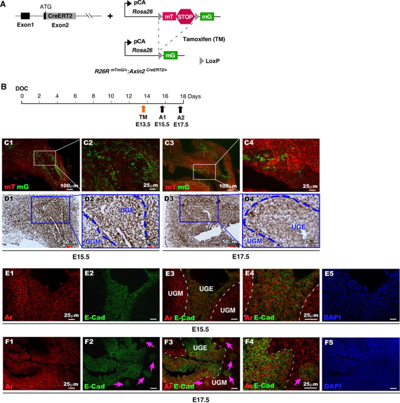
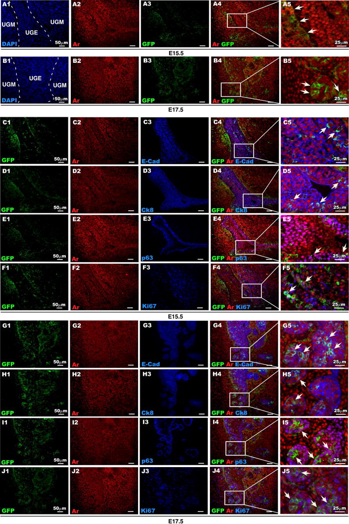
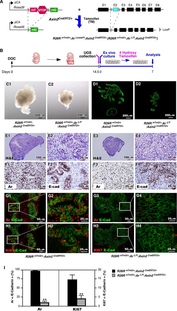
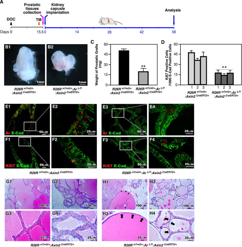
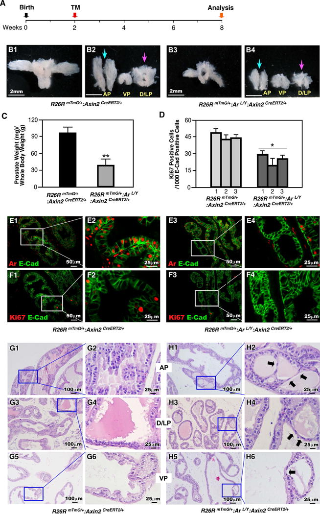
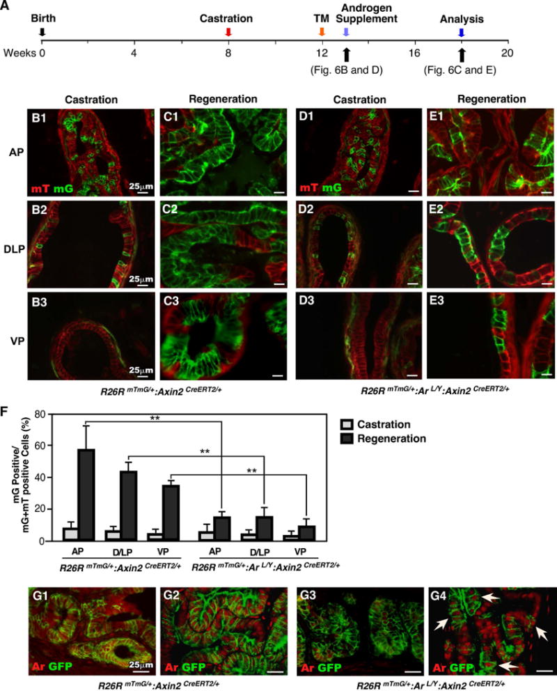
Androgen signaling is essential for prostate development, morphogenesis, and regeneration. Emerging evidence indicates that Wnt/β-catenin signaling also contributes to prostate development specifically through regulation of cell fate determination. Prostatic Axin2-expressing cells are able to respond to Wnt signals and possess the progenitor properties to regenerate prostatic epithelium. Despite critical roles of both signaling pathways, the biological significance of androgen receptor (AR) in Axin2-expressing/Wnt-responsive cells remains largely unexplored. In this study, we investigated this important question using a series of newly generated mouse models. Deletion of Ar in embryonic Axin2-expressing cells impaired early prostate development in both ex vivo and tissue implantation experiments. When Ar expression was deleted in prostatic Axin2-expressing cells at pre-puberty stages, it results in smaller and underdeveloped prostates. A subpopulation of Axin2 expressing cells in prostate epithelium is resistant to castration and, following androgen supplementation, is capable to expand to prostatic luminal cells. Deletion of Ar in these Axin2-expressing cells reduces their regenerative ability. These lines of evidence demonstrate an indispensable role for the Ar in Wnt-responsive cells during the course of prostate development, morphogenesis, and regeneration, which also imply an underlying interaction between the androgen and Wnt signaling pathways in the mouse prostate. Stem Cells 2018;36:891-902.

Keywords: Androgen receptor; Prostate development; Wnt signaling; β-Catenin.


Conflict of interest statement

DISCLOSURE OF POTENTIAL CONFLICTS of INTEREST: The authors indicate no potential conflicts of interests.

Figures

Figure 1
Figure 1. Temporal analysis of Axin2 and Ar expression during early development of mouse prostate
(A): Illustration of transgenic alleles present in R26RmTmG/+:Axin2CreERT2/+ mice. (B): Schematic depicting experimental timeline for labeling and analyzing UGS tissues during embryonic stage. (C): mTomato (red) or mGFP (green) expression in frozen tissue sections of male UGS from E15.5 and E17.5. (D): Ar expression (brown) in male UGS tissues from both E15.5 and E17.5. (E-F): Localization of Ar (red) and E-cadherin (green) in male UGS tissues isolated at E15.5 and E17.5.
Figure 2
Figure 2. Expression of Ar in Axin2-expressing cells during early development of mouse prostate
(A-B): Ar (red) and GFP (green) expression in male UGS isolated at E15.5 and E17.5. (C-F): Colocalization of Ar (red) and GFP (green) with E-cad, CK8, p63 or Ki67 (blue) in UGS tissue isolated at E15.5. (G-J): Colocalization of Ar (red) and GFP (green) with E-cad, CK8, p63 or Ki67 (blue) in UGS tissue isolated at E17.5.
Figure 3
Figure 3. Ar is required in Axin2-expressing cells during prostatic bud formation of ex vivo cultured UGS
(A): Illustration of floxed alleles present in R26RmTmG/+:Arf/Y+:Axin2CreERT2/+mice. (B): Diagram depicting experimental timeline for ex vivo culture of UGS tissues. (C): Gross images of ex vivo cultured UGS tissues isolated from R26RmTmG/+:Axin2CreERT2/+ or R26RmTmG/+:Arf/Y+:Axin2CreERT2/+ mice. (D): E-cadherin expression and localization on sections of ex vivo cultured UGS tissues. (E): H&E stained sections of ex vivo cultured UGS tissues. (F): Ar or E-cadherin (brown) expression pattern in sections of UGS tissues following 7 days of ex vivo culture. (G-H): Colocalization of E-cadherin (green) with Ar or Ki67 (red) in UGS cultured for 7 days ex vivo. (I): Percentage of Ar-expressing cells or Ki67-expressing cells present in urogenital epithelium. ** P<0.01.
Figure 4
Figure 4. Deletion of Ar in Axin2 expressing cells during embryonic stage impairs prostate gland formation in renal capsule of SCID mice
(A): Schematic demonstrating experimental timeline including activation of Axin2CreER, UGS collection, renal capsule transplantation and analysis. (B): Gross images of xenografts from UGS tissue isolated from R26RmTmG/+:Axin2CreERT2/+ or R26RmTmG/+:Arf/Y:Axin2CreERT2/+ mice. (C): Graphical representation of xenograft weight from grown from mice of each genotype. ** P<0.01. (D): Quantification of Ki67 positive cells in the epithelial cellsfrom xenografts of each genotype. ** P<0.01. (E-F): Colocalization of E-cadherin (green) with Ar or Ki67 (red) in R26RmTmG/+:Axin2CreERT2/+ and R26RmTmG/+:Arf/Y:Axin2CreERT2/+ xenografts. (G-H): Representative H&E staining of tissue sections from xenografts of each genotype.
Figure 5
Figure 5. Ar is required in prepubescent Axin2-expressing cells for prostate development
(A): Illustration depicting experimental timeline of Axin2CreER activation (TM injection) and tissue analysis. (B): Gross images of prostates isolated at P56 from R26RmTmG/+:Axin2CreERT2/+ or R26RmTmG/+:Arf/Y:Axin2CreERT2/+ mice. (C): Graphical representation of the ratio of prostate wet weight/body weight. ** P<0.01. (D): Quantification of Ki67 positive cells per 1000 epithelial cells (E-cadherin positive) from prostate tissues of each genotype. * P<0.05. (E-F): Colocalization of E-cadherin (green) with Ar or Ki67 (red) in prostates isolated from R26RmTmG/+:Axin2CreERT2/+ or R26RmTmG/+:Arf/Y:Axin2CreERT2/+ mice. (G-H): H&E staining of tissue sections from prostates of each genotype.
Figure 6
Figure 6. Ar is required for the regenerative potential of castration-resistant Axin2-expressing cells
(A): Diagram of experimental design describing timelines for castration, activation of Axin2CreER (TM injection), androgen supplementation (regeneration) and analysis. (B-E): mTmG analysis of both castrated and regenerated prostate tissues from mice of different genotypes. (F): Graphical quantification of the ratio of mG positive cells per total cells in different lobes of both castrated and regenerated prostates. ** P<0.01. (G) Colocalization of GFP (green) and Ar (red) in frozen sections of both castrated and regenerated prostate tissues from R26RmTmG/+:Axin2CreERT2/+ and R26RmTmG/+:Arf/Y:Axin2CreERT2/+ mice.

References

    1. Balk SP. Androgen receptor as a target in androgen-independent prostate cancer. Urology. 2002;60:132–138. discussion 138-139. - PubMed
    1. Gelmann EP. Molecular biology of the androgen receptor. J Clin Oncol. 2002;20:3001–3015. - PubMed
    1. Zhou ZX, Wong CI, Sar M, et al. The androgen receptor: an overview. Recent Progress Hormone Research. 1994;49:249–274. - PubMed
    1. Cunha GR, Donjacour AA, Cooke PS, et al. The endocrinology and developmental biology of the prostate. Endocr Rev. 1987;8:338–362. - PubMed
    1. Sugimura Y, Cunha GR, Donjacour AA. Morphogenesis of ductal networks in the mouse prostate. Biol Reprod. 1986;34:961–971. - PubMed

Publication types

Substances