Highly Elastic Biodegradable Single-Network Hydrogel for Cell Printing
- PMID: 29451384
- PMCID: PMC5876623
- DOI: 10.1021/acsami.8b01294
Highly Elastic Biodegradable Single-Network Hydrogel for Cell Printing
Abstract
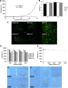
Cell printing is becoming a common technique to fabricate cellularized printed scaffold for biomedical application. There are still significant challenges in soft tissue bioprinting using hydrogels, which requires live cells inside the hydrogels. Moreover, the resilient mechanical properties from hydrogels are also required to mechanically mimic the native soft tissues. Herein, we developed a visible-light cross-linked, single-network, biodegradable hydrogel with high elasticity and flexibility for cell printing, which is different from previous highly elastic hydrogel with double-network and two components. The single-network hydrogel using only one stimulus (visible light) to trigger gelation can greatly simplify the cell printing process. The obtained hydrogels possessed high elasticity, and their mechanical properties can be tuned to match various native soft tissues. The hydrogels had good cell compatibility to support fibroblast growth in vitro. Various human cells were bioprinted with the hydrogels to form cell-gel constructs, in which the cells exhibited high viability after 7 days of culture. Complex patterns were printed by the hydrogels, suggesting the hydrogel feasibility for cell printing. We believe that this highly elastic, single-network hydrogel can be simply printed with different cell types, and it may provide a new material platform and a new way of thinking for hydrogel-based bioprinting research.
Keywords: biodegradable hydrogel; cell printing; elasticity; single network; tissue regeneration.
Conflict of interest statement
The authors declare no competing financial interest.
Figures


Similar articles
-
Bioprinted anisotropic scaffolds with fast stress relaxation bioink for engineering 3D skeletal muscle and repairing volumetric muscle loss.Acta Biomater. 2023 Jan 15;156:21-36. doi: 10.1016/j.actbio.2022.08.037. Epub 2022 Aug 21. Acta Biomater. 2023. PMID: 36002128
-
3D printing of a tough double-network hydrogel and its use as a scaffold to construct a tissue-like hydrogel composite.J Mater Chem B. 2022 Jan 19;10(3):468-476. doi: 10.1039/d1tb02465e. J Mater Chem B. 2022. PMID: 34982091
-
3D bioprinting of urethra with PCL/PLCL blend and dual autologous cells in fibrin hydrogel: An in vitro evaluation of biomimetic mechanical property and cell growth environment.Acta Biomater. 2017 Mar 1;50:154-164. doi: 10.1016/j.actbio.2016.12.008. Epub 2016 Dec 8. Acta Biomater. 2017. PMID: 27940192
-
Exploiting the role of nanoparticles for use in hydrogel-based bioprinting applications: concept, design, and recent advances.Biomater Sci. 2021 Sep 28;9(19):6337-6354. doi: 10.1039/d1bm00605c. Biomater Sci. 2021. PMID: 34397056 Review.
-
Recent advances in high-strength and elastic hydrogels for 3D printing in biomedical applications.Acta Biomater. 2019 Sep 1;95:50-59. doi: 10.1016/j.actbio.2019.05.032. Epub 2019 May 22. Acta Biomater. 2019. PMID: 31125728 Free PMC article. Review.
Cited by
-
Extrusion 3D Printing of Polymeric Materials with Advanced Properties.Adv Sci (Weinh). 2020 Aug 5;7(17):2001379. doi: 10.1002/advs.202001379. eCollection 2020 Sep. Adv Sci (Weinh). 2020. PMID: 32999820 Free PMC article. Review.
-
Bioprinting 101: Design, Fabrication, and Evaluation of Cell-Laden 3D Bioprinted Scaffolds.Tissue Eng Part A. 2020 Mar;26(5-6):318-338. doi: 10.1089/ten.TEA.2019.0298. Tissue Eng Part A. 2020. PMID: 32079490 Free PMC article.
-
Fabrication of Photo-Crosslinkable Poly(Trimethylene Carbonate)/Polycaprolactone Nanofibrous Scaffolds for Tendon Regeneration.Int J Nanomedicine. 2020 Aug 25;15:6373-6383. doi: 10.2147/IJN.S246966. eCollection 2020. Int J Nanomedicine. 2020. PMID: 32904686 Free PMC article.
-
Block Copolymers in 3D/4D Printing: Advances and Applications as Biomaterials.Polymers (Basel). 2023 Jan 8;15(2):322. doi: 10.3390/polym15020322. Polymers (Basel). 2023. PMID: 36679203 Free PMC article. Review.
-
Silk Fibroin Bioink for 3D Printing in Tissue Regeneration: Controlled Release of MSC extracellular Vesicles.Pharmaceutics. 2023 Jan 22;15(2):383. doi: 10.3390/pharmaceutics15020383. Pharmaceutics. 2023. PMID: 36839705 Free PMC article.
References
-
- Lin K.-F.; He S.; Song Y.; Wang C.-M.; Gao Y.; Li J.-Q.; Tang P.; Wang Z.; Bi L.; Pei G.-X. Low-Temperature Additive Manufacturing of Biomimic Three-Dimensional Hydroxyapatite/Collagen Scaffolds for Bone Regeneration. ACS Appl. Mater. Interfaces 2016, 8, 6905–6916. 10.1021/acsami.6b00815. - DOI - PubMed
-
- Yu Y.; Hua S.; Yang M.; Fu Z.; Teng S.; Niu K.; Zhao Q.; Yi C. Fabrication and Characterization of electrospinning/3D Printing Bone Tissue Engineering Scaffold. RSC Adv. 2016, 6, 110557–110565. 10.1039/C6RA17718B. - DOI
-
- Lee S. J.; Lee D.; Yoon T. R.; Kim H. K.; Jo H. H.; Park J. S.; Lee J. H.; Kim W. D.; Kwon I. K.; Park S. A. Surface Modification of 3D-Printed Porous Scaffolds via Mussel-Inspired Polydopamine and Effective Immobilization of rhBMP-2 to Promote Osteogenic Differentiation for Bone Tissue Engineering. Acta Biomater. 2016, 40, 182–191. 10.1016/j.actbio.2016.02.006. - DOI - PubMed
MeSH terms
Substances
Grants and funding
LinkOut - more resources
Full Text Sources
Other Literature Sources

