

Assessment of MRI findings and clinical symptoms in patients with temporomandibular joint disorders
- PMID: 29451403
- PMCID: PMC5991762
- DOI: 10.1259/dmfr.20170412
Assessment of MRI findings and clinical symptoms in patients with temporomandibular joint disorders
Abstract
Objectives: To investigate the correlations among various temporomandibular joint (TMJ) findings on MRI and the relationships between MRI findings and symptoms.
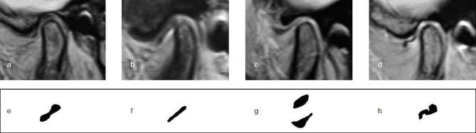
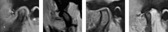
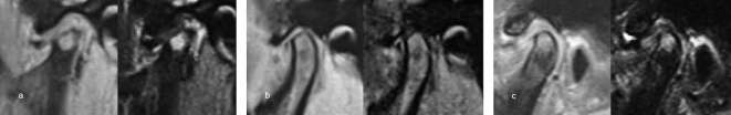
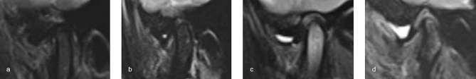
Methods: 425 patients (850 TMJs) with temporomandibular joint disorders (TMDs) who underwent MRI were enrolled. Oblique sagittal proton density-weighted and T2 weighted images in open- and closed-mouth positions were evaluated. MRI findings included disc configuration, disc position, condylar morphology, bone marrow pattern, and joint effusion. Symptoms included TMJ pain, TMJ noise, and limitation of mouth opening. For statistical analyses, Spearman's rank correlation coefficient and logistic regression analysis were applied.
Results: Folded disc, disc displacement without reduction (DDWOR), and osteophytes had significant negative correlations with other normal MRI findings (p < 0.01). DDWOR and marrow edema were associated with TMJ pain. Conversely, osteophytes [odds ratio (OR): 0.52; 95% CI (0.30-0.90)] and combination-type condylar degeneration [OR: 0.45; 95% CI (0.24-0.83)] were associated with decreased risk of TMJ pain. Condylar flattening was positively associated with TMJ noise [OR: 5.25; 95% CI (1.44-19.07)] and negatively associated with limitation of mouth opening [OR: 0.34; 95% CI (0.11-0.99)]. High-grade joint effusion was significantly associated with TMJ pain and noise.
Conclusions: DDWOR and high-grade joint effusion (an indicator of inflammation in the articular cavity) were associated with TMD symptoms. This finding suggests that treatment strategy for DDWOR and decreasing inflammation might lessen clinical TMD symptoms. Condylar degeneration was not associated with indicators of inflammation or TMJ symptoms. These results suggest that patients with TMD symptoms should undergo initial MRI to allow rapid selection of appropriate therapies.
Figures
References
-
- Arayasantiparb R, Tsuchimochi M, Mitrirattanakul S. Transformation of temporomandibular joint disc configuration in internal derangement patients using magnetic resonance imaging. Oral Science International 2012; 9: 43–8. doi: 10.1016/S1348-8643(12)00025-0 - DOI
MeSH terms
LinkOut - more resources
Full Text Sources
Other Literature Sources
Medical
