. 2018 Jun;18(6):1070-1080.
doi: 10.1016/j.spinee.2018.02.007. Epub 2018 Feb 13.

Dental pulp stem cell-derived chondrogenic cells demonstrate differential cell motility in type I and type II collagen hydrogels

Affiliations

Dental pulp stem cell-derived chondrogenic cells demonstrate differential cell motility in type I and type II collagen hydrogels

Li Yao et al. Spine J. 2018 Jun.

Abstract

Background context: Advances in the development of biomaterials and stem cell therapy provide a promising approach to regenerating degenerated discs. The normal nucleus pulposus (NP) cells exhibit similar phenotype to chondrocytes. Because dental pulp stem cells (DPSCs) can be differentiated into chondrogenic cells, the DPSCs and DPSCs-derived chondrogenic cells encapsulated in type I and type II collagen hydrogels can potentially be transplanted into degenerated NP to repair damaged tissue. The motility of transplanted cells is critical because the cells need to migrate away from the hydrogels containing the cells of high density and disperse through the NP tissue after implantation.

Purpose: The purpose of this study was to determine the motility of DPSC and DPSC-derived chondrogenic cells in type I and type II collagen hydrogels.

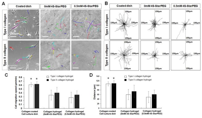
Study design/setting: The time lapse imaging that recorded cell migration was analyzed to quantify the cell migration velocity and distance.

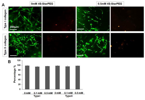
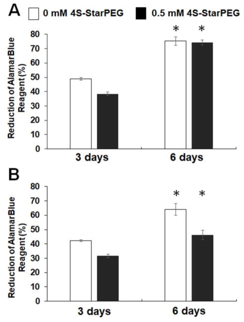
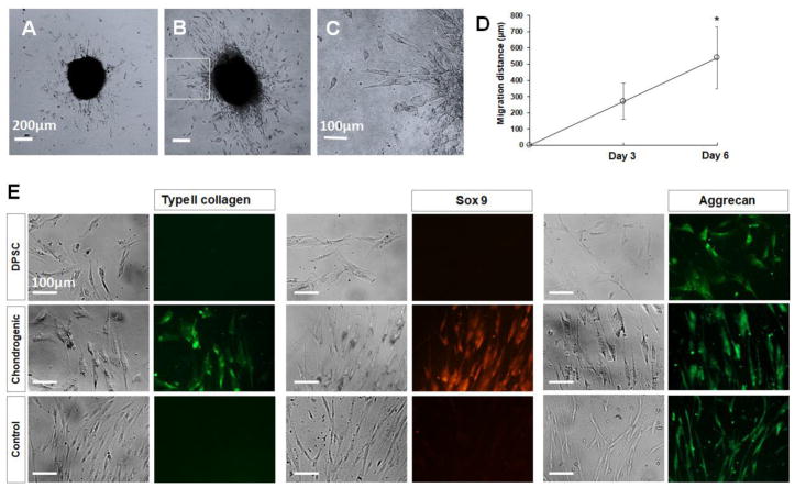
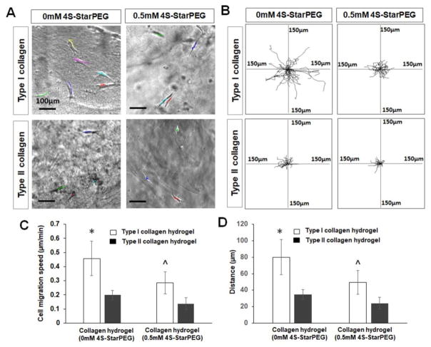
Methods: The cell viability of DPSCs in native or poly(ethylene glycol) ether tetrasuccinimidyl glutarate (4S-StarPEG)-crosslinked type I and type II collagen hydrogels was determined using LIVE/DEAD cell viability assay and AlamarBlue assay. DPSCs were differentiated into chondrogenic cells. The migration of DPSCs and DPSC-derived chondrogenic cells in these hydrogels was recorded using a time lapse imaging system. This study was funded by the Regional Institute on Aging and Wichita Medical Research and Education Foundation, and the authors declare no competing interest.

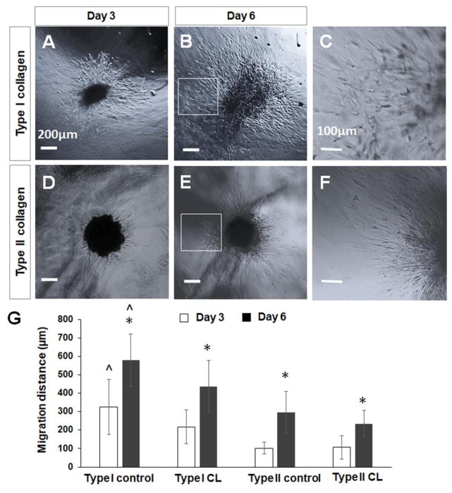
Result: DPSCs showed high cell viability in non-crosslinked and crosslinked collagen hydrogels. DPSCs migrated in collagen hydrogels, and the cell migration speed was not significantly different in either type I collagen or type II collagen hydrogels. The migration speed of DPSC-derived chondrogenic cells was higher in type I collagen hydrogel than in type II collagen hydrogel. Crosslinking of type I collagen with 4S-StarPEG significantly reduced the cell migration speed of DPSC-derived chondrogenic cells.

Conclusions: After implantation of collagen hydrogels encapsulating DPSCs or DPSC-derived chondrogenic cells, the cells can potentially migrate from the hydrogels and migrate into the NP tissue. This study also explored the differential cell motility of DPSCs and DPSC-derived chondrogenic cells in these collagen hydrogels.

Keywords: Chondrogenic cell; Collagen; Dental pulp stem cells; Migration; Nucleus pulposus; Regeneration.


Figures

Figure 1
Figure 1
LIVE/DEAD® cell viability assay for DPSCs grown in collagen hydrogels: (A) Most cells grown in non-crosslinked and 4S-StarPEG crosslinked collagen hydrogels exhibiting high cell viability. Scale bar: 200 μm. The images of same field were taken to show the green or red fluorescence – labeled cells. Live cells labeled with calcein AM (green). Dead cells labeled with ethidium homodimer-1 (red). (B) Percentage of live cells in collagen hydrogels as determined by LIVE/DEAD® cell assay.
Figure 2
Figure 2
AlamarBlue® assay for DPSCs grown in hydrogels for 6 days demonstrating cell proliferation in these hydrogels. *, p < 0.05, compared with cell culturing for 3 days.
Figure 3
Figure 3
Migration of DPSCs in hydrogels. (A) DPSC cells migrated in cell culture dish or in hydrogels (each line indicates one migration track of a cell). Scale bar: 100 μm. (B) Cell migration paths determined by video monitor tracings (position of all cells at t = 0 min represented by origin position (center of frame), with migratory track of each cell at 3 hours plotted as single line on graph; each axis arm represents 150 μm of translocation distance). (C) Quantification of DPSC migration speed in hydrogels. (D) Quantification of cell migration distance in hydrogels. *, p < 0.05, compared with DPSC cell migration in corresponding type I or type II collagen hydrogel.
Figure 4
Figure 4
Migration and differentiation of DPSC-derived chondrogenic cells. (A–D) DPSC-derived chondrogenic cells migrated out of cell pellet after culturing on collagen-coated cell culture dish. Scale bar Figures A and B: 200 μm. Scale bar Figure C: 100 μm. (E) Cells that migrated out of the pellets labeled with anti-type II collagen, anti-sox 9, and anti-aggrecan antibodies. Scale bar: 100 μm.
Figure 5
Figure 5
DMMB assay of level of GAGs in cell culture medium produced by chondrogenic pellets and control pellets. *, p < 0.05, compared with corresponding control pellets after pellet culturing for 1 week and 2 weeks.
Figure 6
Figure 6
Migration of DPSC-derived chondrogenic cells of cell pellets in collagen hydrogels. (A–F) DPSC-derived chondrogenic cells migrated into collagen hydrogels from cell pellets. (A, B) Cell migration in type I collagen hydrogel. (C, D) Cell migration in type II collagen hydrogel. Scale bar: 200 μm. (C) Magnified images of inset indicated in (B). (F) Magnified images of inset indicated in (E). Scale bar: 100 μm. (G) Quantification of cell migration distance in collagen hydrogels from cell pellets. *, p < 0.05, compared with cell migration in corresponding collagen hydrogels after cell culturing for 3 days. ^, p < 0.05, compared with cell migration in corresponding type II collagen hydrogels and crosslinked type I and type II collagen hydrogels after cell culturing for 3 days and 6 days. CL, crosslinked collagen.
Figure 7
Figure 7
Migration of DPSC-derived chondrogenic cells in hydrogels. (A) Cells migrated in collagen hydrogels (each line indicates one migration track of a cell). Scale bar: 100 μm. (B) Cell migration paths determined by video monitor tracings (position of all cells at t = 0 minute represented by origin position (center of frame), with migratory track of each cell at 3 hours plotted as single line on graph; each axis arm represents 150 μm of translocation distance). (C) Quantification of cell migration speed in hydrogels. (D) Quantification of cell migration distance in hydrogels. *, p < 0.05, compared with cell migration in type II collagen hydrogel and crosslinked type I and type II collagen hydrogels. ^, p < 0.05, compared with cell migration crosslinked type II collagen hydrogel.

Similar articles

Cited by

References

    1. Hunter CJ, Matyas JR, Duncan NA. The notochordal cell in the nucleus pulposus: a review in the context of tissue engineering. Tissue Eng. 2003;9:667–677. doi: 10.1089/107632703768247368. - DOI - PubMed
    1. Sakai D. Future perspectives of cell-based therapy for intervertebral disc disease. Eur Spine J. 2008;17(Suppl 4):452–458. doi: 10.1007/s00586-008-0743-5. - DOI - PMC - PubMed
    1. Kandel R, Roberts S, Urban JP. Tissue engineering and the intervertebral disc: the challenges. Eur Spine J. 2008;17(Suppl 4):480–491. doi: 10.1007/s00586-008-0746-2. - DOI - PMC - PubMed
    1. Mern DS, Beierfuss A, Thome C, Hegewald AA. Enhancing human nucleus pulposus cells for biological treatment approaches of degenerative intervertebral disc diseases: a systematic review. J Tissue Eng Regen Med. 2012 doi: 10.1002/term.1583. - DOI - PubMed
    1. Gan Y, Li P, Wang L, Mo X, Song L, Xu Y, Zhao C, Ouyang B, Tu B, Luo L, Zhu L, Dong S, Li F, Zhou Q. An interpenetrating network-strengthened and toughened hydrogel that supports cell-based nucleus pulposus regeneration. Biomaterials. 2017 Aug;136:12–28. doi: 10.1016/j.biomaterials.2017.05.017. - DOI - PubMed

Publication types