. 2018 Mar;118(3):502-513.
doi: 10.1055/s-0038-1627453. Epub 2018 Feb 16.

Suppressive Role of Tissue Factor Pathway Inhibitor-α in Platelet-Dependent Fibrin Formation under Flow Is Restricted to Low Procoagulant Strength

Affiliations

Suppressive Role of Tissue Factor Pathway Inhibitor-α in Platelet-Dependent Fibrin Formation under Flow Is Restricted to Low Procoagulant Strength

Stella Thomassen et al. Thromb Haemost. 2018 Mar.

Abstract

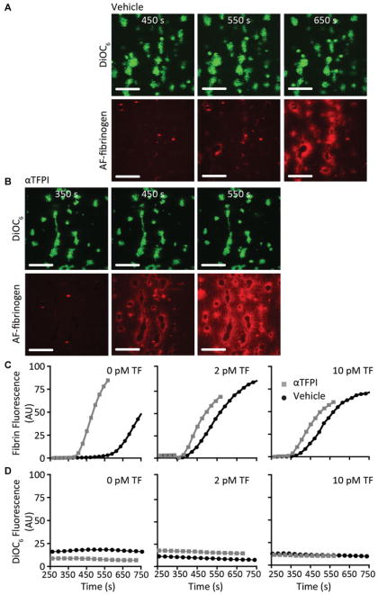
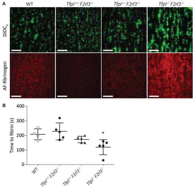
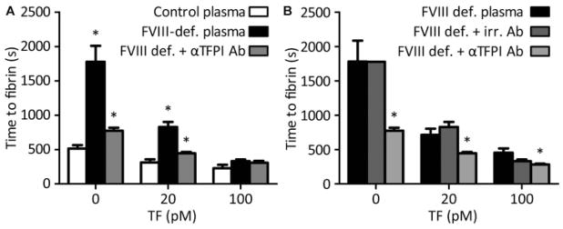
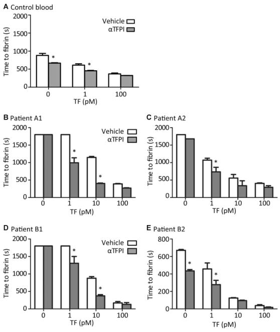
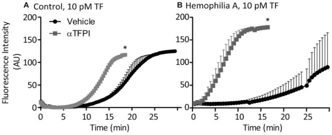
Tissue factor pathway inhibitor-alpha (TFPI-α) is a Kunitz-type serine protease inhibitor, which suppresses coagulation by inhibiting the tissue factor (TF)/factor VIIa complex as well as factor Xa. In static plasma-phospholipid systems, TFPI-α thus suppresses both factor Xa and thrombin generation. In this article, we used a microfluidics approach to investigate how TFPI-α regulates fibrin clot formation in platelet thrombi at low wall shear rate. We therefore hypothesized that the anticoagulant effect of TFPI-α in plasma is a function of the local procoagulant strength-defined as the magnitude of thrombin generation under flow, due to local activities of TF/factor VIIa and factor Xa. To test this hypothesis, we modulated local coagulation by microspot coating of flow channels with 0 to 100 pM TF/collagen, or by using blood from patients with haemophilia A or B. For blood or plasma from healthy subjects, blocking of TFPI-α enhanced fibrin formation, extending from a platelet thrombus, under flow only at <2 pM coated TF. This enhancement was paralleled by an increased thrombin generation. For mouse plasma, genetic deficiency in TFPI enhanced fibrin formation under flow also at 0 pM TF microspots. On the other hand, using blood from haemophilia A or B patients, TFPI-α antagonism markedly enhanced fibrin formation at microspots with up to 100 pM coated TF. We conclude that, under flow, TFPI-α is capable to antagonize fibrin formation in a manner dependent on and restricted by local TF/factor VIIa and factor Xa activities.


Conflict of interest statement

A.E.M. receives research grant support from Novo Nordisk. The remaining authors state that they have no conflict of interest.

Figures

Fig. 1
Fig. 1
TFPI-α inhibition enhances fibrin formation on platelet thrombi under flow at low TF density. Platelet thrombi were generated by perfusion of human blood over three consecutive microspots of collagen with increasing amounts of 0, 2 and 10 pM coated TF (4 minutes at 1,000 s−1). Platelet thrombi were superfused at shear rate of 150 s−1 with recalcified normal plasma (vehicle or TFPIα inhibited); fibrin formation from the platelet thrombi was determined from recorded images. Shown are representative brightfield images, captured at indicated times after start of perfusion with vehicle-treated (A) or TFPI-α-inhibited (B) plasma (no TF, bar = 25 μm). (C) Time to fibrin formation at microspots with consecutively 0, 2 and 10 pM TF. Means ± SEM, n = 4–5; *p < 0.05 versus vehicle; NS, not significant.
Fig. 2
Fig. 2
Effect of TFPI-α inhibition on kinetics of platelet-dependent fibrin formation under flow. Platelet thrombi were generated by perfusion of DiOC6-labeled human blood over microspots with collagen and indicated amounts of TF, as for Fig. 1. Platelet thrombi were superfused with recalcified normal plasma containing AF647 fibrinogen. Shown are representative confocal fluorescence images (no TF, time points indicated) after perfusion with vehicle-treated plasma (A) or TFPI-α-inhibited plasma (B). Bars = 25 μm. Time traces of fibrin formation (C) and platelet adhesion (D) (fluorescence image analysis) at platelet thrombi for microspots with 0, 2 and 10 pM TF (n = 3).
Fig. 3
Fig. 3
Deficiency in murine TFPI enhances fibrin formation on platelet thrombi under flow. Wild-type murine platelet thrombi were generated whole blood (DiOC6-labeled) perfusion over collagen microspots. Recalcified plasmas containing AF647-fibrinogen from wild type, Tfpi+/+ F2rl3−/−, Tfp+;/− F2rl3−/− or Tfpi−/− F2rl3−/− mice were post-perfused at low shear rate (150 s−1), while DiOC6 and AF647 fluorescence was monitored by confocal microscopy. (A) Representative DiOC6 and AF647-fibrin images after 200 s of flow (bars = 25 μm). (B) Times to first fibrin formation per genotype, measured by image subtraction analysis. Means ± SEM, n = 4–5; *p < 0.05 versus wild type.
Fig. 4
Fig. 4
TFPI-α inhibition restores fibrin formation of haemophiliac plasma on platelet thrombi at higher TF density. Platelet thrombi were generated by blood perfusion over microspots with collagen and increasing amounts of 0, 20 and 100 pM coated TF. Thrombi were superfused with indicated recalcified plasmas, and time to fibrin formation was recorded as for Fig. 1. (A) Perfusion with control or factor (F)VIII-deficient plasma, and depletion of TFPI-α as indicated. (B) Perfusion with factor VIII–deficient plasma, treated with irrelevant (irr.) control antibody or anti-TFPI antibodies. Means ± SEM, n = 4–7; *p < 0.05 versus control plasma (A) or FVIII-deficient plasma (B).
Fig. 5
Fig. 5
TFPI-α inhibition normalizes platelet-dependent fibrin formation in haemophiliac blood. Platelet thrombi were generated by blood perfusion over microspots of collagen with indicated concentrations of TF; blood samples were treated with vehicle or anti-TFPI antibodies. Time to first fibrin formation was recorded per microspot during blood perfusion at 150 s−1. Presented are times to fibrin formation. (A) Blood from control subjects. (B, C) Blood from haemophilia A patients A1 (2% factor VIII) and A2 (<1% with 3.8 BU factor VIII inhibitor). (D, E) Blood from haemophilia B patients B1 (5% factor IX) and B2 (7% factor IX). Means ± SEM, n = 3–4; *p < 0.05 versus vehicle.
Fig. 6
Fig. 6
Effect of TFPI-α inhibition on whole blood thrombin generation on platelet thrombi under stasis. Thrombi with procoagulant platelets were generated by whole blood perfusion over 10 pM TF/collagen microspots. Blood samples were pretreated with vehicle or anti-TFPI antibodies, and supplemented with Z-GGR-AMC (0.5 mM). After 4 minutes, the fluorescence accumulation of cleaved thrombin substrate was recorded under stasis to assess for thrombin generation. Shown are representative thrombin generation curves with blood from control subjects (A) or haemophilia A patients (B). Means ± SEM (n = 3), *p < 0.05 versus vehicle.

Similar articles

Cited by

References

    1. Salemink I, Franssen J, Willems GM, Hemker HC, Lindhout T. Inhibition of tissue factor-factor VIIa-catalyzed factor X activation by factor Xa-tissue factor pathway inhibitor. A rotating disc study on the effect of phospholipid membrane composition. J Biol Chem. 1999;274(40):28225–28232. - PubMed
    1. Adams M. Tissue factor pathway inhibitor: new insights into an old inhibitor. Semin Thromb Hemost. 2012;38(02):129–134. - PubMed
    1. Broze GJ, Jr, Girard TJ. Tissue factor pathway inhibitor: structure-function. Front Biosci (Landmark Ed) 2012;17:262–280. - PMC - PubMed
    1. Winckers K, ten Cate H, Hackeng TM. The role of tissue factor pathway inhibitor in atherosclerosis and arterial thrombosis. Blood Rev. 2013;27(03):119–132. - PubMed
    1. Maroney SA, Ellery PE, Wood JP, Ferrel JP, Martinez ND, Mast AE. Comparison of the inhibitory activities of human tissue factor pathway inhibitor (TFPI)α and TFPIβ. J Thromb Haemost. 2013;11(05):911–918. - PMC - PubMed

MeSH terms